Right surface
Description:
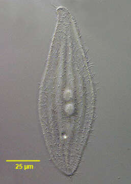
Portrait (right surface) of the rhabdophorine ciliate, Siroloxophyllum utriculariae (Penard,1922) Foissner,1995. Siroloxophyllum was erected as a new genus based on the "adoral bulge" which encircles almost the entire circumference of the cell, a single dorsolateral brush kinety and morphologically distinct right and left Dorsolateral kineties different from other somatic kineties. The strongly laterally compressed cell is lancet shaped in outline. The cell is slightly contractile and highly flexible. The rounded anterior end is curved dorsally. The posterior is bluntly tapered. The flat right side bears 13-20 longitudinal kineties. The left side has 3-8 prominent longitudinal ridges bearing short cilia. The slit-like cytostome is located along the anteroventral edge. A hyaline band containing long rod shaped extrusomes borders the cell. There is a distinctive structure which looks like a helix or twisted cord bordering the entire edge of the cell except for a short length of the anterior dorsal end. The perpendicularly arranged peripheral extrusomes appear to anchor their exterior ends in this structure. There are two contractile vacuoles. The central macronucleus is bipartite. The inconspicuous micronucleus is located between the two parts of the macronucleus (not seen here). S. utriculariae swims slowly, gliding gracefully over the substrate. Differentiated from the similar L. helus by absence of trichocyst warts along the dorsal surface. Most easily confused with Amphileptus species which lack the distinctive bordering cord-like structure described above above except for a short part of the anterior ventral surface and also have an anterior kinetal suture (spica) on the right surface. May also be confused with Litonotus species which usually have a single contractile vacuole and extrusomes limited to only part of the ventral surface. Collected from a freshwater dredge pond near Boise, Idaho October 2004. DIC.
Included On The Following Pages:
- Life (creatures)
- Cellular (cellular organisms)
- Eukaryota (eukaryotes)
- SAR (Stramenopiles, Alveolates, Rhizaria)
- Alveolata (alveolates)
- Ciliophora (ciliates)
- Intramacronucleata
- Litostomatea
- Haptoria
- Pleurostomatida
- Litonotidae
- Siroloxophyllum
- Siroloxophyllum utricularium
This image is not featured in any collections.
Source Information
- license
- cc-by-nc
- author
- William Bourland
- provider
- micro*scope
- original
- original media file
- visit source
- partner site
- micro*scope
- ID