portrait
Description:
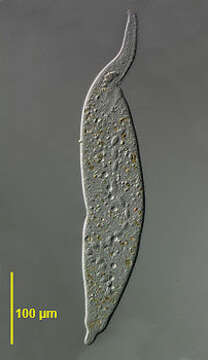
Portrait of the large ciliate, Monilicaryon monilatus (Stokes, 1886) Jankowski, 1967. This cell is slightly compressed. Similar in overall appearance to Dileptus anser. M. monilatus differs by having a shorter proboscis relative to the length of the body (1/3 to 1/4) and by lacking the row of obliquely oriented closely spaced kinetids on the ventral aspect of the left side of the proboscis (this feature requires demonstration by DIC or protargol staining). M. monilatus has two single files of kinetids extending from either side of the oral aperture anteriorly along the ventral aspect of the proboscis separated by a strip bearing extrusomes (See Foissner W., Berger H and Kohmann F. Taxonomische und ökologische Revision der Ciliaten des Saprobiensystems- Band IV: Gymnostomatea, Loxodes, Suctoria. Informationsberichte Bayer. Landesamtes für Wasserwirtschaft. 1/95:185-202, 1995). The moniliform macronucleus is seen here. There are 20-40 small dorsal contractile vacuoles each emptying through a single excretory pore. There is often a larger contractile vacuole at the base of the tail. Collected from a freshwater pond near Boise, Idaho. DIC.
Included On The Following Pages:
- Life (creatures)
- Cellular (cellular organisms)
- Eukaryota (eukaryotes)
- SAR (Stramenopiles, Alveolates, Rhizaria)
- Alveolata (alveolates)
- Ciliophora (ciliates)
- Intramacronucleata
- Litostomatea
- Haptoria
- Haptorida
- Dileptida
- Dileptidae
- Monilicaryon
- Monilicaryon monilatum
This image is not featured in any collections.
Source Information
- license
- cc-by-nc
- author
- William Bourland
- provider
- micro*scope
- original
- original media file
- visit source
- partner site
- micro*scope
- ID