Image of thismia
Description:
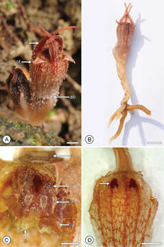
Figure 2.Flower structure in Thismia hongkongensis sp. nov. A Mature flower, showing outer tepals (ot), inner tepals (it) and abscission zone (ab) at the base of the perianth tube. B Entire plant (S.S. Mar 1, HK). C Perianth tube with annulus (a), following removal of the proximal face of the tube, exposing pendent stamens with filament (f), thecae (th), connective (c) and lateral appendage (la) (S.S. Mar 2, HK). D Inner face of perianth tube, showing network patterning and putative nectaries (arrowed) (S.S. Mar 2, HK). Scale bars: A, D = 2 mm; B = 5 mm; C = 1 mm. Photos: A, B S.S. Mar; C, D R.M.K. Saunders.
Included On The Following Pages:
- Life (creatures)
- Cellular (cellular organisms)
- Eukaryota (eukaryotes)
- Archaeplastida (plants)
- Chloroplastida (green plants)
- Streptophyta
- Embryophytes
- Tracheophyta (ferns)
- Spermatophytes (seed plants)
- Angiosperms (Dicotyledons)
- Monocots (Monocotyledons)
- Dioscoreales
- Burmanniaceae
- Thismia (thismia)
- Thismia hongkongensis
This image is not featured in any collections.
Source Information
- license
- cc-by-3.0
- copyright
- Shek Shing Mar, Richard M.K. Saunders
- bibliographic citation
- Mar S, Saunders R (2015) Thismia hongkongensis (Thismiaceae): a new mycoheterotrophic species from Hong Kong, China, with observations on floral visitors and seed dispersal PhytoKeys (46): 21–33
- original
- original media file
- visit source
- partner site
- Phytokeys
- ID