-
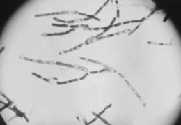
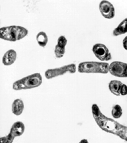
This photomicrograph depicted a number of Gram-positive, endospore-forming Bacillus anthracis bacteria. B. anthracis is the pathologic microorganism responsible for the disease anthrax, an acute infectious disease, which most commonly occurs in wild and domestic vertebrates (cattle, sheep, goats, camels, antelopes, and other herbivores), but it can also occur in humans when they are exposed to infected animals, or tissue from infected animals.Created: 1982
-
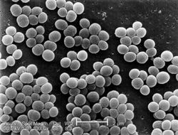
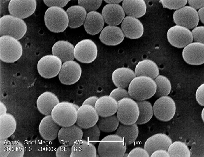
Under a very high magnification of 20,000x, this scanning electron micrograph (SEM) shows a strain of Staphylococcus aureus bacteria taken from a vancomycin intermediate resistant culture (VISA).Under SEM, one can not tell the difference between bacteria that are susceptible, or multidrug resistant, but with transmission electron microscopy (TEM), at least with VISA isolates one can see a thickening in the cell wall that may attribute to their reduced susceptibility to vancomycin . See PHIL 11157 for a colorized version of this image. VISA and VRSA are specific types of antimicrobial-resistant staph bacteria. While most staph bacteria are susceptible to the antimicrobial agent vancomycin some have developed resistance. VISA and VRSA cannot be successfully treated with vancomycin because these organisms are no longer susceptibile to vancomycin. However, to date, all VISA and VRSA isolates have been susceptible to other Food and Drug Administration (FDA) approved drugs.Created: 2001
-
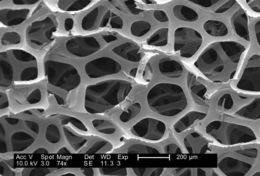
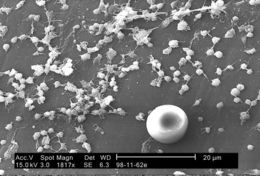
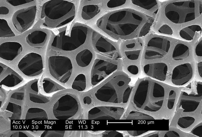
Under a low magnification of 76x, this 2007 scanning electron micrograph (SEM) depicted the fibrous configuration of a dry macrofoam sponge swabs. This swab, as well as three other materials, including polyester (see PHIL 9735), rayon (see PHIL 9734) and cotton (see PHIL 9732, and 9733), were scanned for a CDC study involving their efficiency in recovery of Bacillus anthracis bacterial spores from steel coupons that had been inoculated with a spore suspension of known concentration. See PHIL 9736, 9737, 9738, 9749, 9750, and 9751, for other views of this material. The article discussing the description of this swab material analysis, and the analytical results was published in Emerging Infectious Diseases, Vol. 10, No. 6, June, 2004, and was entitled, Swab Materials and Bacillus anthracis Spore Recovery from Nonporous Surfaces. A link to this article is found below.Created: 2007
-
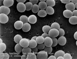
Under a high magnification of 10,000x, this scanning electron micrograph (SEM) shows a strain of Staphylococcus aureus bacteria taken from a vancomycin intermediate resistant culture (VISA).Under SEM, one can not tell the difference between bacteria that are susceptible, or multidrug resistant, but with transmission electron microscopy (TEM), at least with VISA isolates one can see a thickening in the cell wall that may attribute to their reduced susceptibility to vancomycin . See PHIL 11155 for a colorized version of this image.Created: 2001
-
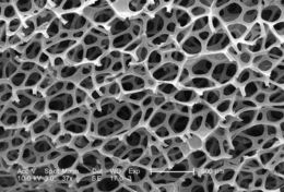
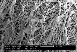
Under a low magnification of 74x, this 2007 scanning electron micrograph (SEM) depicted the fibrous configuration of a dry macrofoam sponge swabs. This swab, as well as three other materials, including polyester (see PHIL 9735), rayon (see PHIL 9734) and cotton (see PHIL 9732, and 9733), were scanned for a CDC study involving their efficiency in recovery of Bacillus anthracis bacterial spores from steel coupons that had been inoculated with a spore suspension of known concentration. See PHIL 9736, 9737, 9738, 9749, 9751, an 9752, for other views of this material. The article discussing the description of this swab material analysis, and the analytical results was published in Emerging Infectious Diseases, Vol. 10, No. 6, June, 2004, and was entitled, Swab Materials and Bacillus anthracis Spore Recovery from Nonporous Surfaces. A link to this article is found below.Created: 2007
-
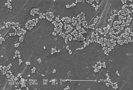
This 2005 scanning electron micrograph (SEM) depicted numerous clumps of methicillin-resistant Staphylococcus aureus bacteria, commonly referred to by the acronym, MRSA; Magnified 2381x. Recently recognized outbreaks, or clusters of MRSA in community settings have been associated with strains that have some unique microbiologic and genetic properties, compared with the traditional hospital-based MRSA strains, which suggests some biologic properties, e.g., virulence factors like toxins, may allow the community strains to spread more easily, or cause more skin disease. A common strain named USA300-0114 has caused many such outbreaks in the United States. See PHIL 10048 for a colorized version of this micrograph.Created: 2005
-
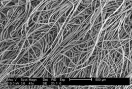
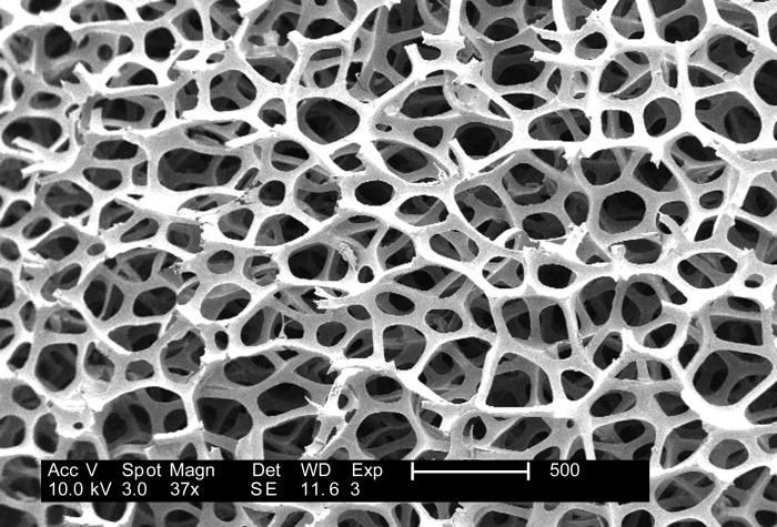
Under a low magnification of 37x, this 2007 scanning electron micrograph (SEM) depicted the fibrous configuration of a dry macrofoam sponge swabs. This swab, as well as three other materials, including polyester (see PHIL 9735), rayon (see PHIL 9734) and cotton (see PHIL 9732, and 9733), were scanned for a CDC study involving their efficiency in recovery of Bacillus anthracis bacterial spores from steel coupons that had been inoculated with a spore suspension of known concentration. See PHIL 9736, 9737, 9738, 9750, 9751, an 9752, for other views of this material. The article discussing the description of this swab material analysis, and the analytical results was published in Emerging Infectious Diseases, Vol. 10, No. 6, June, 2004, and was entitled, Swab Materials and Bacillus anthracis Spore Recovery from Nonporous Surfaces. A link to this article is found below.Created: 2007
-
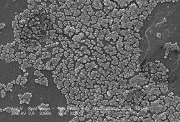
This 2005 scanning electron micrograph (SEM) depicted numerous clumps of methicillin-resistant Staphylococcus aureus bacteria, commonly referred to by the acronym, MRSA; Magnified 2390x. Recently recognized outbreaks, or clusters of MRSA in community settings have been associated with strains that have some unique microbiologic and genetic properties, compared with the traditional hospital-based MRSA strains, which suggests some biologic properties, e.g., virulence factors like toxins, may allow the community strains to spread more easily, or cause more skin disease. A common strain named USA300-0114 has caused many such outbreaks in the United States. Please see PHIL 10047 for a colorized version of this image.Created: 2005
-
Under a low magnification of 188x, this 2007 scanning electron micrograph (SEM) depicted the fibrous configuration of a dry macrofoam sponge swab. This swab, as well as three other materials, including polyester (see PHIL 9735), rayon (see PHIL 9734), and cotton (see PHIL 9732, 9733) were scanned for a CDC study involving their efficiency in recovery of Bacillus anthracis bacterial spores from steel coupons that had been inoculated with a spore suspension of known concentration. See PHIL 9736, and 9737 for other views of this material. The article discussing the description of this swab material analysis, and the analytical results was published in Emerging Infectious Diseases, Vol. 10, No. 6, June, 2004, and was entitled, Swab Materials and Bacillus anthracis Spore Recovery from Nonporous Surfaces. A link to this article is found below.Created: 2007
-
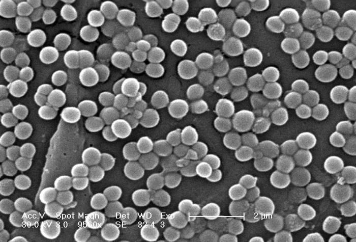
This 2005 scanning electron micrograph (SEM) depicted numerous clumps of methicillin-resistant Staphylococcus aureus bacteria, commonly referred to by the acronym, MRSA; Magnified 9560x.Recently recognized outbreaks, or clusters of MRSA in community settings have been associated with strains that have some unique microbiologic and genetic properties, compared with the traditional hospital-based MRSA strains, which suggests some biologic properties, e.g., virulence factors like toxins, may allow the community strains to spread more easily, or cause more skin disease. A common strain named USA300-0114 has caused many such outbreaks in the United States. See PHIL 10046 for a colorized version of this micrograph.Created: 2005
-
Under a low magnification of 37x, this 2007 scanning electron micrograph (SEM) depicted the fibrous configuration of a dry macrofoam sponge swab. This swab, as well as three other materials, including polyester (see PHIL 9735), rayon (see PHIL 9734), and cotton (see PHIL 9732, 9733) were scanned for a CDC study involving their efficiency in recovery of Bacillus anthracis bacterial spores from steel coupons that had been inoculated with a spore suspension of known concentration. See PHIL 9736, and 9738 for other views of this material. The article discussing the description of this swab material analysis, and the analytical results was published in Emerging Infectious Diseases, Vol. 10, No. 6, June, 2004, and was entitled, Swab Materials and Bacillus anthracis Spore Recovery from Nonporous Surfaces. A link to this article is found below.Created: 2007
-
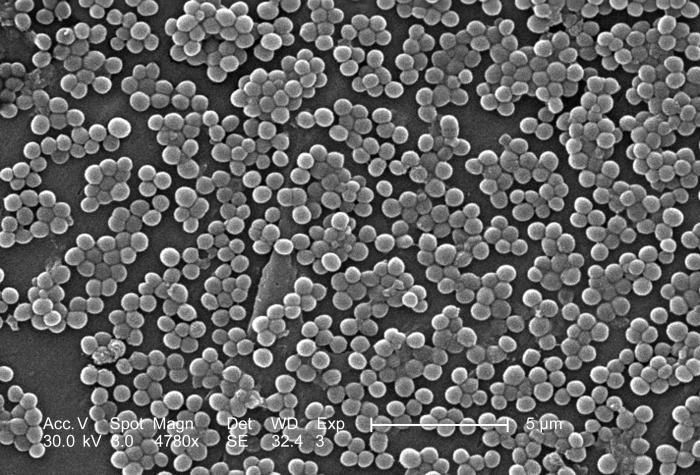
This 2005 colorized scanning electron micrograph (SEM) depicted numerous clumps of methicillin-resistant Staphylococcus aureus bacteria, commonly referred to by the acronym, MRSA; Magnified 4780. Recently recognized outbreaks, or clusters of MRSA in community settings have been associated with strains that have some unique microbiologic and genetic properties, compared with the traditional hospital-based MRSA strains, which suggests some biologic properties, e.g., virulence factors like toxins, may allow the community strains to spread more easily, or cause more skin disease. A common strain named USA300-0114 has caused many such of outbreaks in the United States. See PHIL 10045 for a colorized version of this image.Created:
-
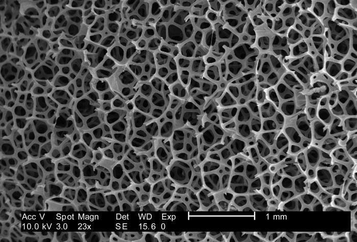
Under a low magnification of 23x, this 2007 scanning electron micrograph (SEM) depicted the fibrous configuration of a dry macrofoam sponge swab. This swab, as well as three other materials, including polyester (see PHIL 9735), rayon (see PHIL 9734), and cotton (see PHIL 9732, 9733) were scanned for a CDC study involving their efficiency in recovery of Bacillus anthracis bacterial spores from steel coupons that had been inoculated with a spore suspension of known concentration. See PHIL 9737, and 9738 for other views of this material. The article discussing the description of this swab material analysis, and the analytical results was published in Emerging Infectious Diseases, Vol. 10, No. 6, June, 2004, and was entitled, Swab Materials and Bacillus anthracis Spore Recovery from Nonporous Surfaces. A link to this article is found below.Created:
-
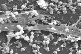
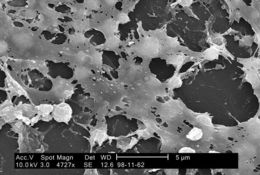
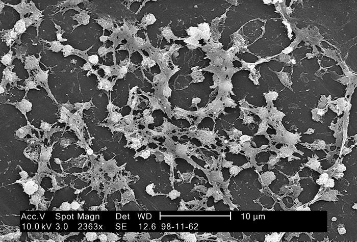
This electron micrograph depicted numbers of Staphylococcus aureus bacteria, which were fond on the luminal surface of an indwelling catheter. A red blood cell (RBD), also known as an erythrocyte is present with its biconcave cytomorphology. Of importance is the sticky-looking substance woven between the round cocci bacteria, which was composed of polysaccharides, and is known as biofilm. This biofilm has been found to protect the bacteria that secrete the substance from attacks by antimicrobial agents such as antibiotics; Magnified 2363x.S. aureus, often referred to simply as "staph," are bacteria commonly carried on the skin, or in the nose of healthy people. Approximately 25% to 30% of the population is colonized, i.e., when bacteria are present, but not causing an infection, in the nose with staph bacteria.Created:
-
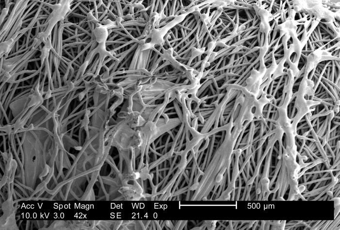
Under a low magnification of 42x, this 2007 scanning electron micrograph (SEM) depicted the fibrous configuration of a plain uninoculated polyester swab. This swab, as well as three other materials, including macrofoam (see PHIL 9736, 9737, 9738), rayon (see PHIL 9734), and cotton (see PHIL 9732, 9733) were scanned for a CDC study involving their efficiency in recovery of Bacillus anthracis bacterial spores from steel coupons that had been inoculated with a spore suspension of known concentration. The article discussing the description of this swab material analysis, and the analytical results was published in Emerging Infectious Diseases, Vol. 10, No. 6, June, 2004, and was entitled, Swab Materials and Bacillus anthracis Spore Recovery from Nonporous Surfaces. A link to this article is found below.Created: 2007
-
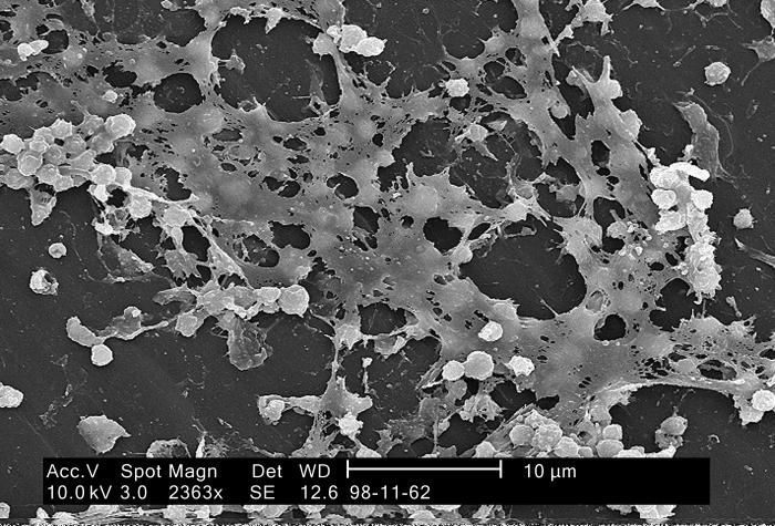
This electron micrograph depicted large numbers of Staphylococcus aureus bacteria, which were fond on the luminal surface of an indwelling catheter. Of importance is the sticky-looking substance woven between the round cocci bacteria, which was composed of polysaccharides, and is known as biofilm. This biofilm has been found to protect the bacteria that secrete the substance from attacks by antimicrobial agents such as antibiotics; Magnified 2363x.S. aureus, often referred to simply as "staph," are bacteria commonly carried on the skin, or in the nose of healthy people. Approximately 25% to 30% of the population is colonized, i.e., when bacteria are present, but not causing an infection, in the nose with staph bacteria.Created: 2005
-
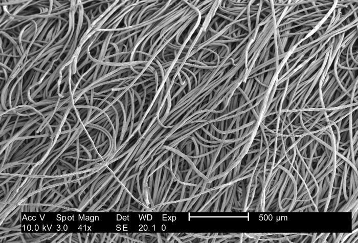
Under a low magnification of 41x, this 2007 scanning electron micrograph (SEM) depicted the fibrous configuration of a plain uninoculated rayon swab. This swab, as well as three other materials, including macrofoam (see PHIL 9736, 9737, 9738), polyester (see PHIL 9735), and cotton (see PHIL 9732, 9733) were scanned for a CDC study involving their efficiency in recovery of Bacillus anthracis bacterial spores from steel coupons that had been inoculated with a spore suspension of known concentration. The article discussing the description of this swab material analysis, and the analytical results was published in Emerging Infectious Diseases, Vol. 10, No. 6, June, 2004, and was entitled, Swab Materials and Bacillus anthracis Spore Recovery from Nonporous Surfaces. A link to this article is found below.Created: 2007
-
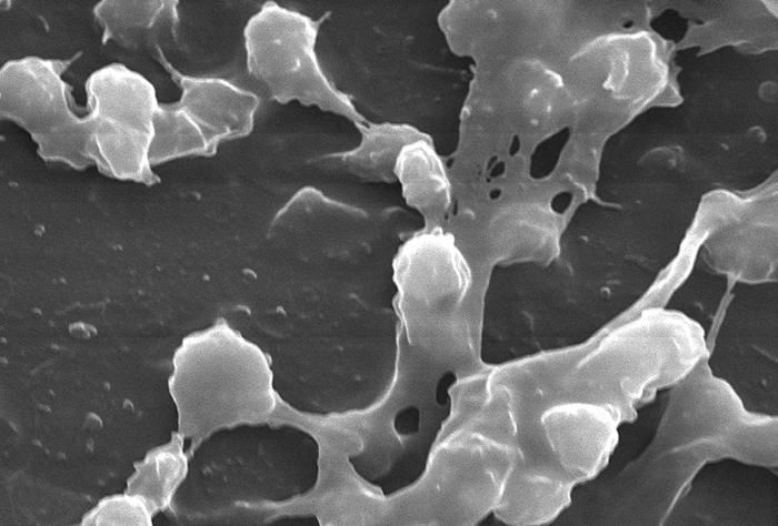
This highly magnified electron micrograph depicted numbers of Staphylococcus aureus bacteria, which were fond on the luminal surface of an indwelling catheter. Of importance is the sticky-looking substance woven between the round cocci bacteria, which was composed of polysaccharides, and is known as biofilm. This biofilm has been found to protect the bacteria that secrete the substance from attacks by antimicrobial agents such as antibiotics; Magnified 2363x.S. aureus, often referred to simply as "staph," are bacteria commonly carried on the skin, or in the nose of healthy people. Approximately 25% to 30% of the population is colonized, i.e., when bacteria are present, but not causing an infection, in the nose with staph bacteria.Created: 2005
-
Under a low magnification of 38x, this 2007 scanning electron micrograph (SEM) depicted the fibrous configuration of a plain uninoculated cotton swab. This swab, as well as three other materials, including macrofoam (see PHIL 9736, 9737, 9738), polyester (see PHIL 9735), and rayon (see PHIL 9734) were scanned for a CDC study involving their efficiency in recovery of Bacillus anthracis bacterial spores from steel coupons that had been inoculated with a spore suspension of known concentration. See PHIL 9732, for another view of this material. The article discussing the description of this swab material analysis, and the analytical results was published in Emerging Infectious Diseases, Vol. 10, No. 6, June, 2004, and was entitled, Swab Materials and Bacillus anthracis Spore Recovery from Nonporous Surfaces. A link to this article is found below.Created: 2007
-
This highly magnified electron micrograph depicted numbers of Staphylococcus aureus bacteria, which were fond on the luminal surface of an indwelling catheter. Of importance is the sticky-looking substance woven between the round cocci bacteria, which was composed of polysaccharides, and is known as biofilm. This biofilm has been found to protect the bacteria that secrete the substance from attacks by antimicrobial agents such as antibiotics; Magnified 2363x.S. aureus, often referred to simply as "staph," are bacteria commonly carried on the skin, or in the nose of healthy people. Approximately 25% to 30% of the population is colonized, i.e., when bacteria are present, but not causing an infection, in the nose with staph bacteria.Created: 2005
-
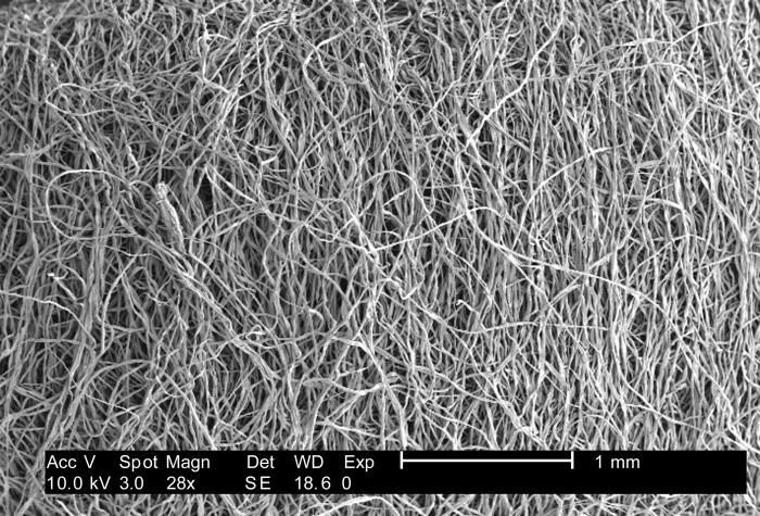
Under a low magnification of 28x, this 2007 scanning electron micrograph (SEM) depicted the fibrous configuration of a plain uninoculated cotton swab. This swab, as well as three other materials, including macrofoam (see PHIL 9736, 9737, 9738), polyester (see PHIL 9735), and rayon (see PHIL 9734) were scanned for a CDC study involving their efficiency in recovery of Bacillus anthracis bacterial spores from steel coupons that had been inoculated with a spore suspension of known concentration. See PHIL 9733, for another view of this material. The article discussing the description of this swab material analysis, and the analytical results was published in Emerging Infectious Diseases, Vol. 10, No. 6, June, 2004, and was entitled, Swab Materials and Bacillus anthracis Spore Recovery from Nonporous Surfaces. A link to this article is found below.Created: 2007
-
This highly magnified electron micrograph depicted numbers of Staphylococcus aureus bacteria, which were fond on the luminal surface of an indwelling catheter. Of importance is the sticky-looking substance woven between the round cocci bacteria, which was composed of polysaccharides, and is known as biofilm. This biofilm has been found to protect the bacteria that secrete the substance from attacks by antimicrobial agents such as antibiotics; Magnified 2363x.S. aureus, often referred to simply as "staph," are bacteria commonly carried on the skin, or in the nose of healthy people. Approximately 25% to 30% of the population is colonized, i.e., when bacteria are present, but not causing an infection, in the nose with staph bacteria.Created: 2005
-
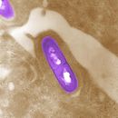
Transmission electron micrograph of Bacillus anthracis.Created: 2001
-
This electron micrograph depicted large numbers of Staphylococcus aureus bacteria, which were fond on the luminal surface of an indwelling catheter. Of importance is the sticky-looking substance woven between the round cocci bacteria, which was composed of polysaccharides, and is known as biofilm. This biofilm has been found to protect the bacteria that secrete the substance from attacks by antimicrobial agents such as antibiotics; Magnified 2363x.S. aureus, often referred to simply as "staph," are bacteria commonly carried on the skin, or in the nose of healthy people. Approximately 25% to 30% of the population is colonized, i.e., when bacteria are present, but not causing an infection, in the nose with staph bacteria.Created: 2005