-
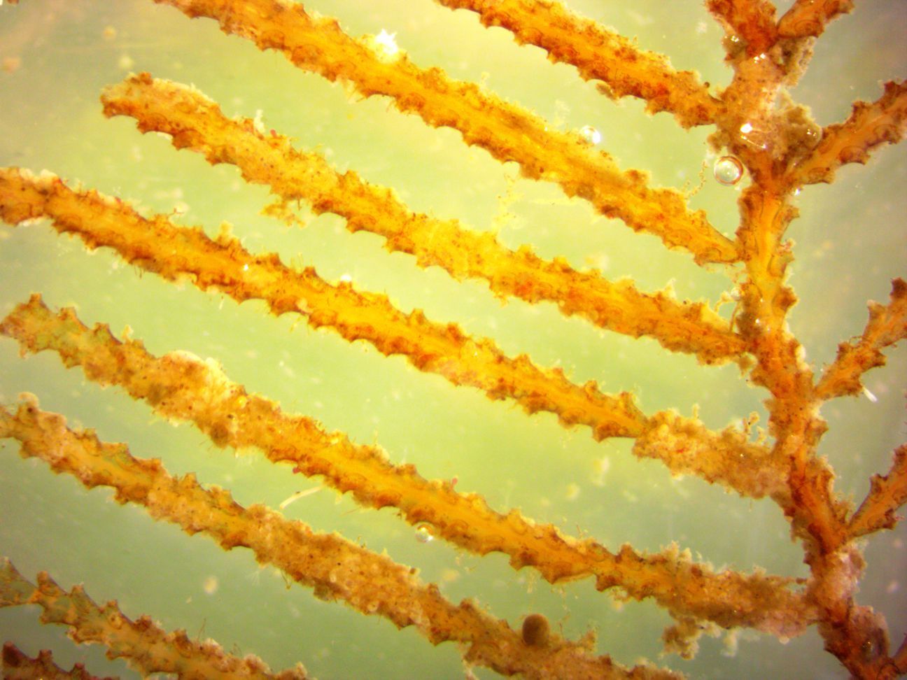
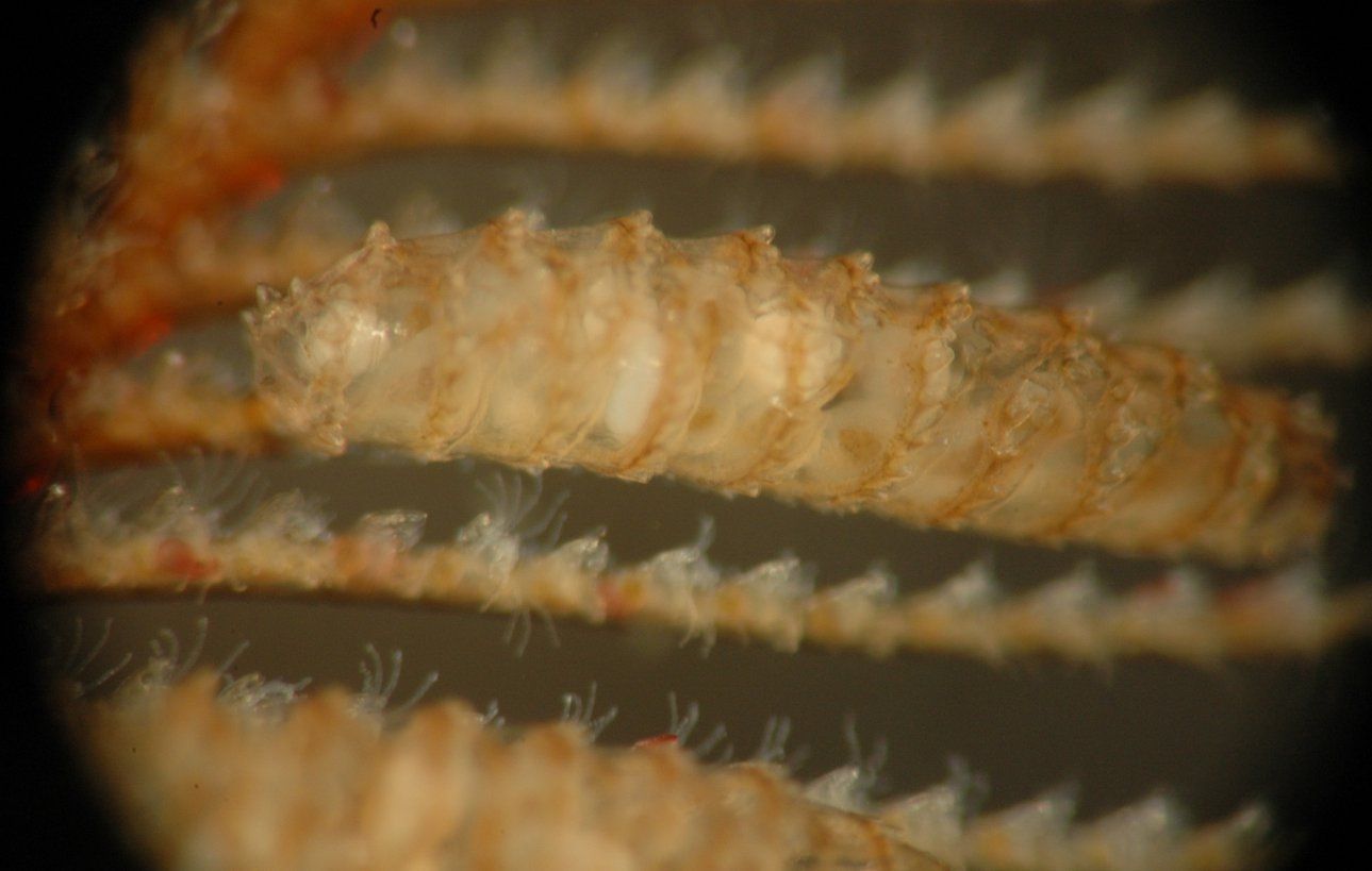
This closer view of the colony shows the polyps (within their hydrothecae) along the two sides of each branch. Notice that the branches are about twice as wide as the hydrothecae are.
-
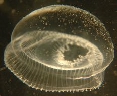
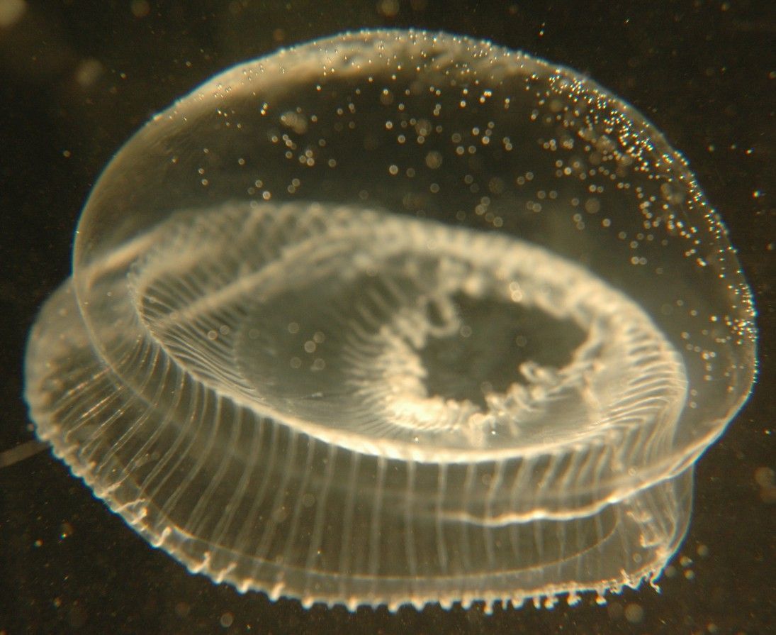
Aequorea victoria, 4 cm diameter, captured in central Rosario Strait July 12, 2007. The broad, open manubrium with frilly lips can be seen through the bell. The tentacles are retracted. (Photo by: Dave Cowles, July 2007)
-
Melicertum octocostatum (Sars, 1835)
-
The individual hydrothecae can be clearly seen in this closer view. The colony is a bit fouled by diatoms and other debris.
-
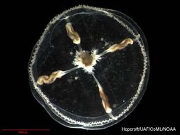
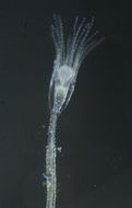
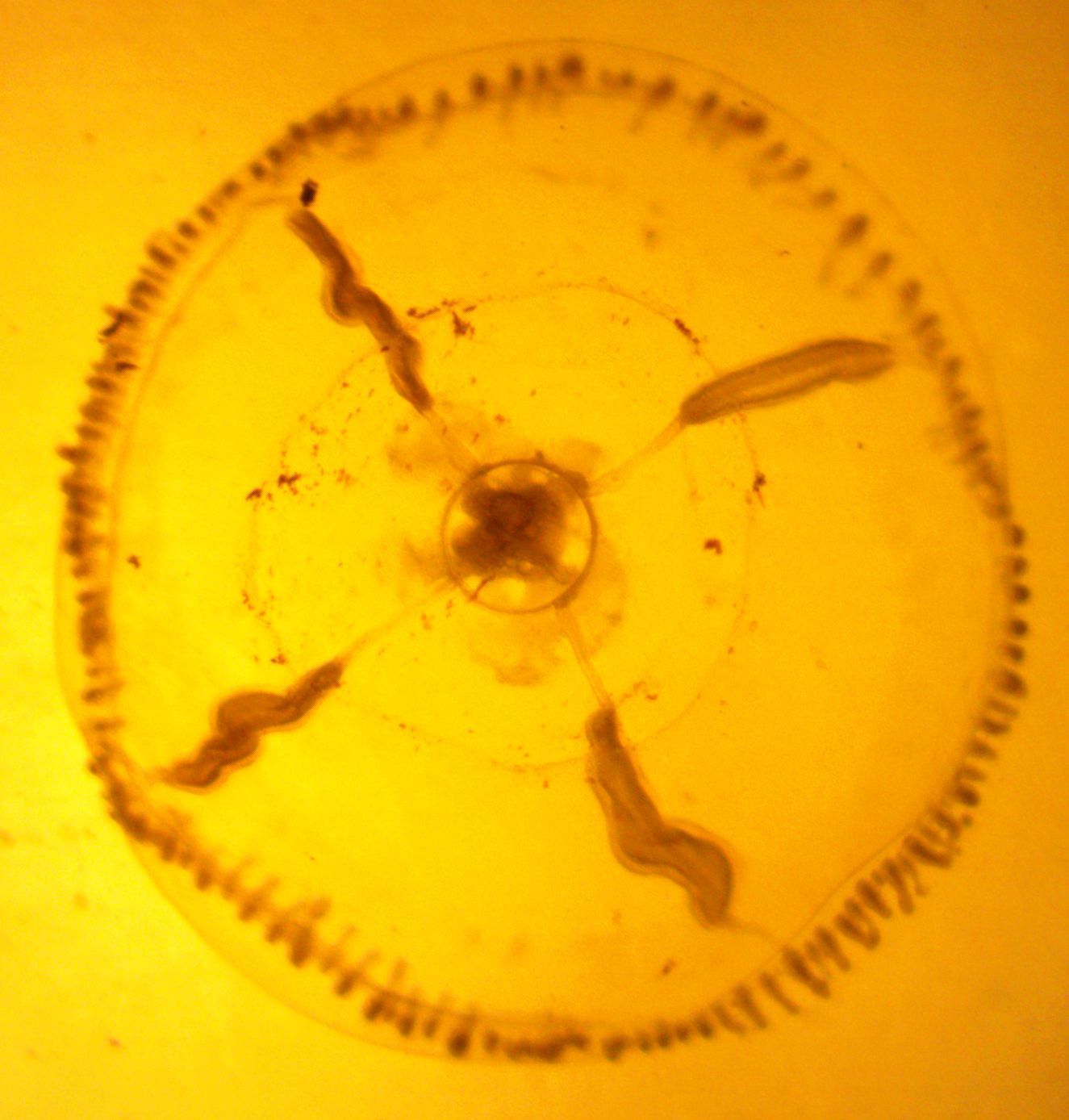
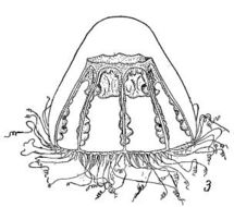
This photo taken in transmitted light highlights many of the features of the hydromedusa.
-
Mitrocomella polydiademata (Romanes, 1876)
-
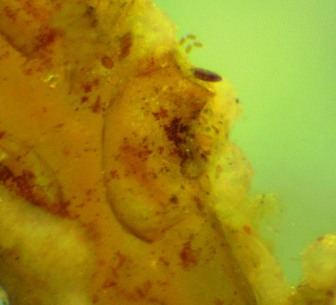
In this photo of an individual hydrotheca, the darker operculum which is attached on the side nearest the stalk can be seen.
-
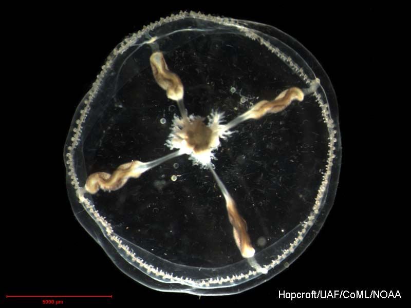
Eutonina indicans, 8 mm diameter, captured on a midwater plankton tow in the Strait of Juan de Fuca (Photo by: Dave Cowles, July 2014)
-
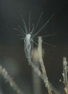
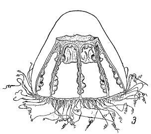
Tiaropsis multicirrata (Sars, 1835)
-
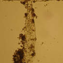
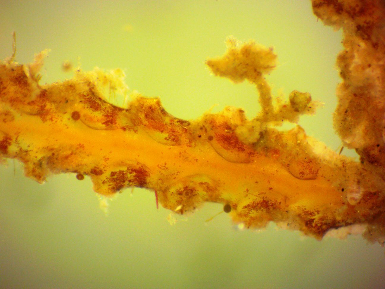
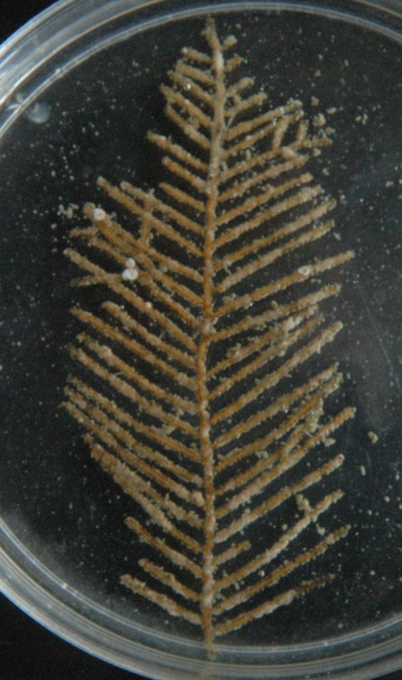
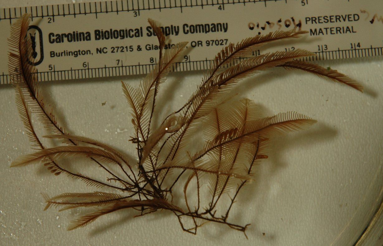
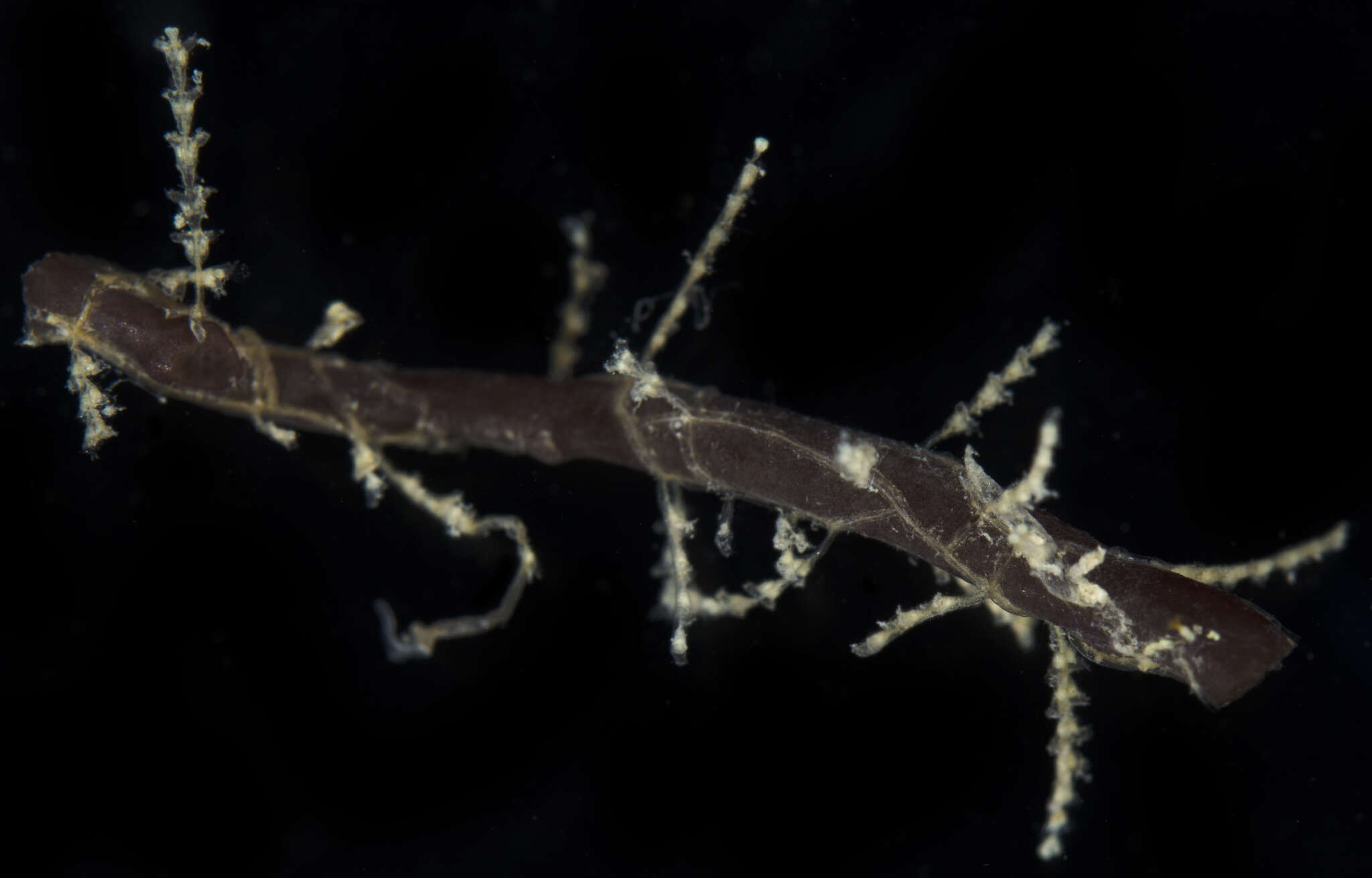
Abietinaria inconstans colony, about 8 cm tall, found at 90 m depth in the San Juan Channel. This colony is heavily encrusted with small bryozoans and tubeworms. (Photo by: Dave Cowles, August 2014)
-
Tiaropsis multicirrata (Sars, 1835)
-
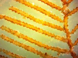
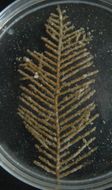
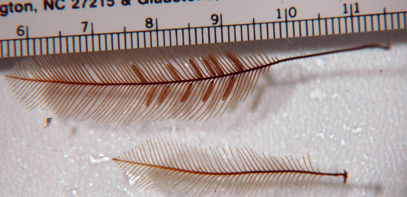
A closer view of two Aglaophenia plumes. The upper plume has corbulae with the reproductive gonophores.
-
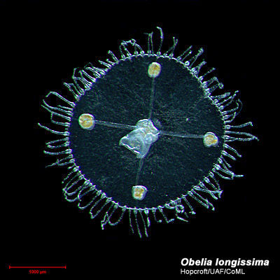
Obelia longissima (Pallas, 1766)
-
This closeup of a plume shows the individual polyps in their hydrothecae (note some polyps are open and actively feeding while others are retracted). The large structures are a string of corbulae with the reproductive gonophores (and captive medusae) inside.
-
Obelia longissima (Pallas, 1766)
-
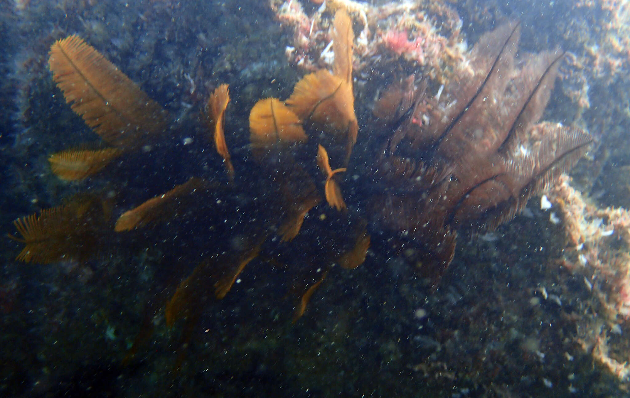
Aglaophenia sp. colony collected at 5 m depth off Sares Head. (Photo by: Dave Cowles, Jule 2006)
-
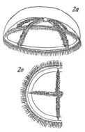
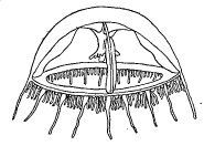
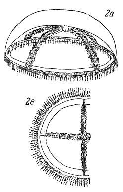
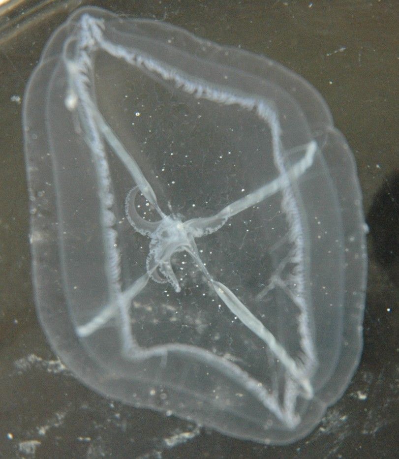
Staurophora mertensii Brandt, 1838
-
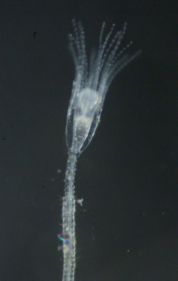
Another indivudual on the same tunicate. Note the diatoms attached to some stalks.
-
Orthopyxis everta, about 2 mm tall, on a tunicate. Note the annulated pedicel and smooth rim to the hydrotheca. (Photo by: Dave Cowles, August 2010 )
-
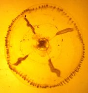
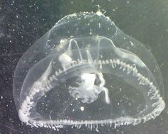
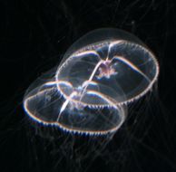
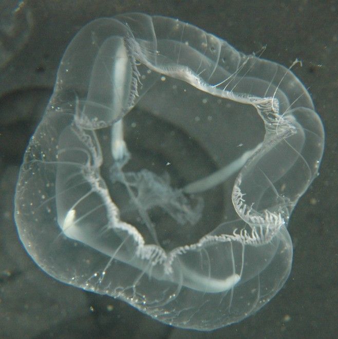
The tentacles are numerous (up to 350), lined up along the margin of the bell (not in clusters), and highly extensible. In this view most of them are highly contracted. When extended, long and shorter ones alternate.
-
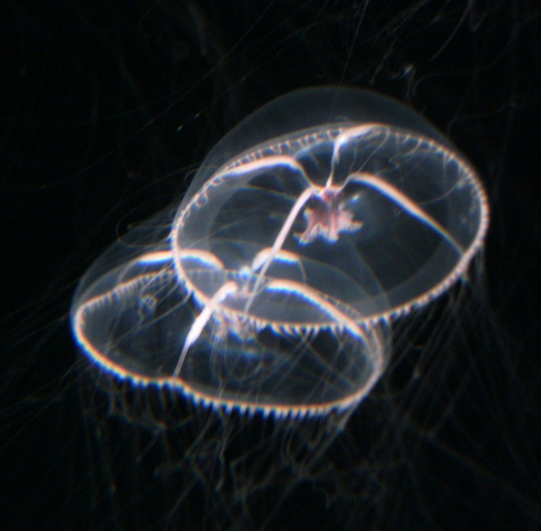
Mitrocoma cellularia in Monterey Bay Aquarium. Photo by Dave Cowles, August 2010
-
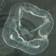
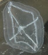
Mitrocoma cellularia from Friday Harbor. Diameter about 4 cm. (Photo by: Dave Cowles, July 2009 )
-
-