Brief Summary
(
Inglês
)
fornecido por IABIN
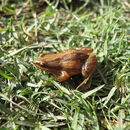
Diagnosis A very small elongate and slender species, characterized by a glandulous skin, with evident longitudinal ridges. Enlarged and flattened glandular patchcs on the sides and a glandular ridge behind mouth joint are present.
- licença
- cc-by-nc-sa-3.0
- direitos autorais
- Museo Nacional de Historia Natural
- autor
- Esteban O. Lavilla
- editor
- Diego Arrieta
Distribution
(
Inglês
)
fornecido por IABIN
Known in the provinces of Buenos Aires and Entre Ríos in Argentina; Rio Grande do Sul and Santa Catarina states in Brazil and Uruguay.
- licença
- cc-by-nc-sa-3.0
- direitos autorais
- Museo Nacional de Historia Natural
- autor
- Esteban O. Lavilla
- editor
- Diego Arrieta
Diagnostic Description
(
Inglês
)
fornecido por IABIN
Adult morphology Snout-vent of about 17-22 mrn in males, 18-24 mrn in females. Head as large as wide; snout rounded slightly protruding, with dorso-lateral nostrils, almost at the tip of the snout. Internarial interval narrower than the interocular distance, which is broader than the upper eyelid. Canthus rostralis blun, rounded; loreal region concave. Maxillary teeth present; no vomerine teeth. Tongue oval, entire and free behind. Eyes rnoderately prominent, laterally located; their diameter sligtly smaller than the length of the snout.Tympanum hardly recognizable. Flat supratympanic fold, reaching behind the axilla. Fingers rounded at the tip, free: subarticuIar and metacarpal tubercles blunt and promineat. Rate of the finger lengths: I-II-IV-II. Forearms wartv, but no antebrachial tubercles. Toes free, slender, subarticular and metatarsal tubercles conical and prominent, A faint tarsal tubercle slightly closer to the inner metatarsal tubercle than to the heel. Tarsal fold wealr but distinct. When hindleg is adpressed, heel goes beyond the shoulder. When the femurs are bent at right angles to body, the tibio-tarsal articulations overlap. Skin glandular on the dorsurn, smooth on the beIlv. Faint and broken longitudinal ridges from eyes backward. An enlarged area latero-posteriorly, from the axilla to groin. A bulky glandular ridge behind the mouth joint. Posterior region of the thighs coarsely glandular. No rounded inguinal spots present. No discoidal fold evident. Dorsal color ground brownish, scattered with darker spots on the back and limbs. Ventrally whitish or yellowish, immaculate. Larval morphology The tadpoles have a depressed body; the body length is about a third of the total length; the body shape is ovoid in dorsal view and the maximum width is placed in the posterior or middle third of the body. In lateral view, the ventral contour of the body is slightly convex, almost flat. The snout is rounded in dorsal and lateral views. The nostrils are round, with a fleshy, slightly elevated marginal rim; they are dorsolaterally positioned, closer to the tip of the snout than to the eyes, and are visible in dorsal, lateral and frontal views. The eyes are large, placed in dorsal position and directed dorsolaterally. The pineal end organ is not evident. The spiracle is single, lateral, sinistral and short; it is placed in the middle third of the body; its inner wall is fused to the body, and its opening is oval, elevated, with a diameter smaller than the tube diameter, and it is visible in lateral and ventral views. The intestinal assa is located approximately at the centre of the ventral region. Neuromasts are not visible. The vent tube has a medial configuration, it is wider next to the body and its opening is round, although it may be subject to folding; it opens medially, but in one case the opening is directed to the right, due to a folding at the origin of the ventral fin. The tail is large, with both fins higher than body height. The dorsal fin originates at the body-tail junction, and the ventral fin origin is attached to the vent tube. Both fins have regularly curved margins. The tail axis is straight, the tip is acute and the miotomes of the tail musculature are conspicuous, especially in the proximal region. The oral disc is anteroventral, small and laterally emarginated. It has a single row of marginal papillae, with a large dorsal gap, and a short ventral gap. The papillae are simple, with rounded tip. The submarginal papillae are absent. The keratinized structures (jaw sheaths and labial teeth) are partially or completely lost in some tadpoles. When present, the upper jaw sheath has a serrated free margin, is laterally convex and flat in the middle part. The lower jaw sheath has a free margin U to V-shaped, is serrated and heavily pigmented. The labial tooth row formula is 2(2)/3(1), the length of P-3 is about 50% of the other rowsÂ? length, and is approximately as long as the ventral gap of marginal papillae. In vivo coloration: Dorsum yellowish brown with dark blotches. Ventrally silver, sometimes transparent. Tail musculature yellowish brown, darker on the upper side, with some iridescent mottling. Fins transparent with dark brown mottling and iridescent spots, mainly in the ventral fin. Iris golden copper. Margin of nostrils darker than the surrounding skin. Some tadpoles have a dorsal light yellowish midline, from the head to the middle of the tail.
- licença
- cc-by-nc-sa-3.0
- direitos autorais
- Museo Nacional de Historia Natural
- autor
- Esteban O. Lavilla
- editor
- Diego Arrieta