-
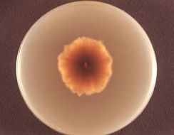
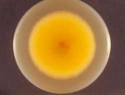
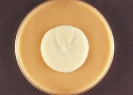
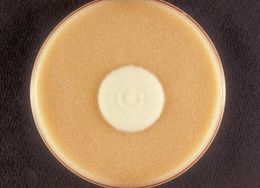
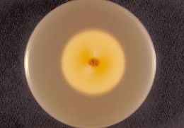
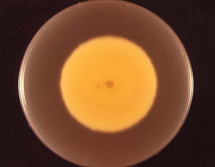
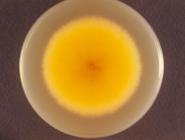
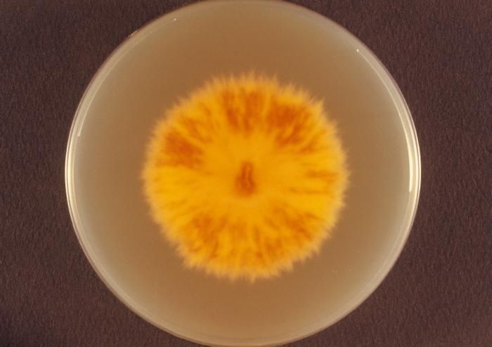
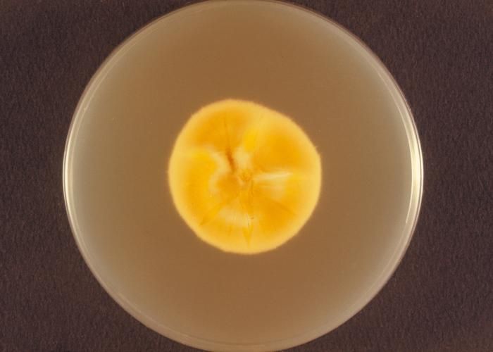
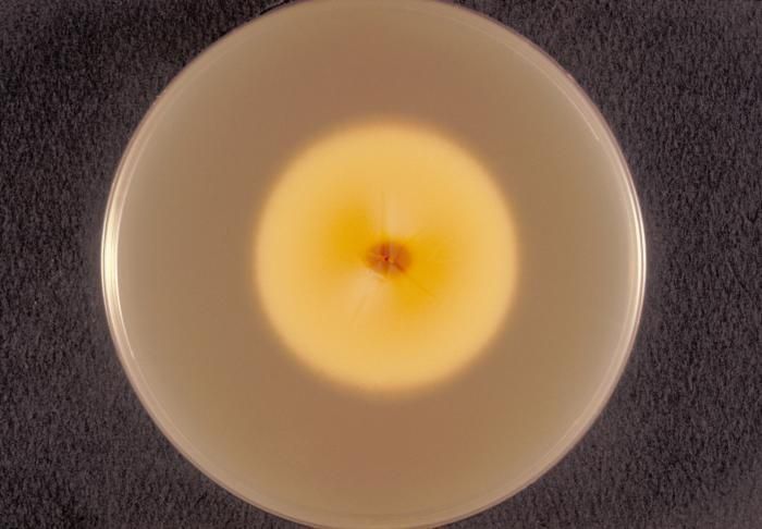
Viewed from the back, i.e., reverse, this image depicted a Petri dish containing Sabouraud's (SAB) dextrose agar, upon which a Microsporum persicolor fungal colony had been cultured. As seen in this reverse view, the colonial coloration can be yellow, or may even be a red-brown. From the front, as depicted in PHIL 10904 and 10906, the colonies can be white, or depending upon the Microsporum sp., may run the gamut, sporting a yellow, beige or cinnamon color, and display a flat, or glabrous, woolly or powdery texture.Created: 1973
-
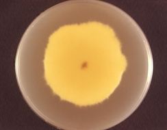
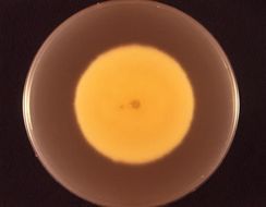
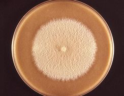
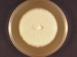
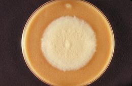
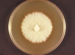
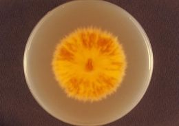
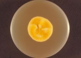
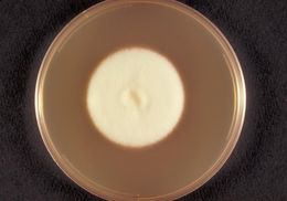
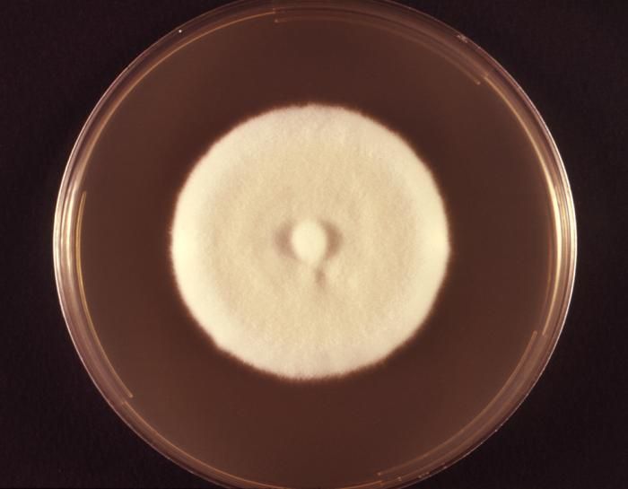
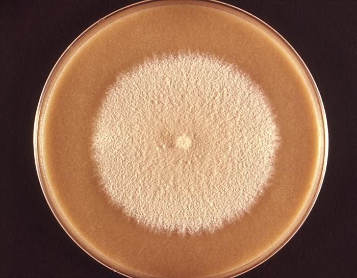
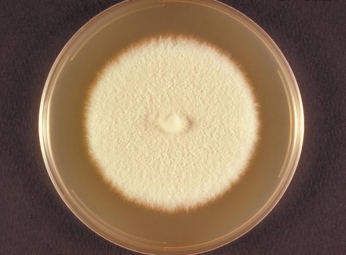
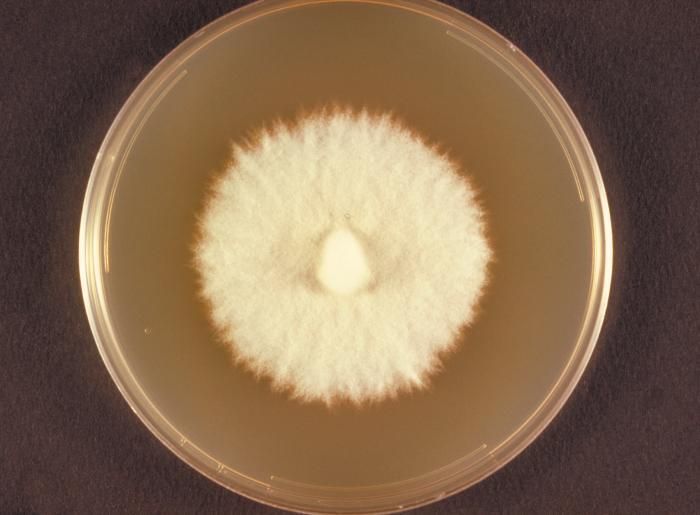
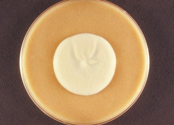
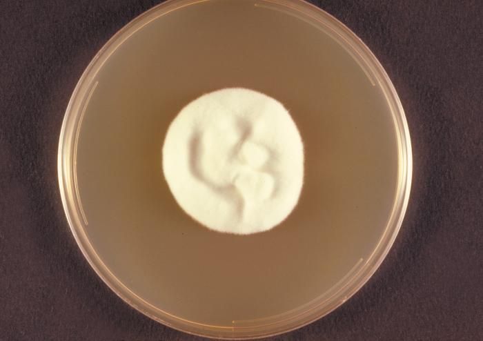
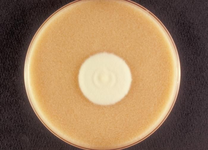
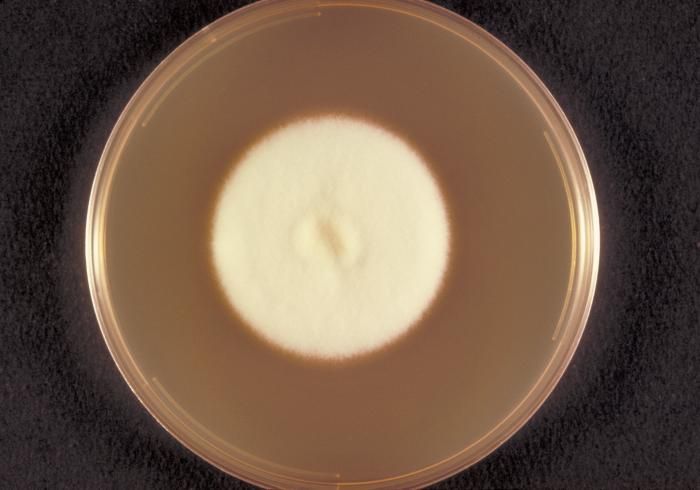
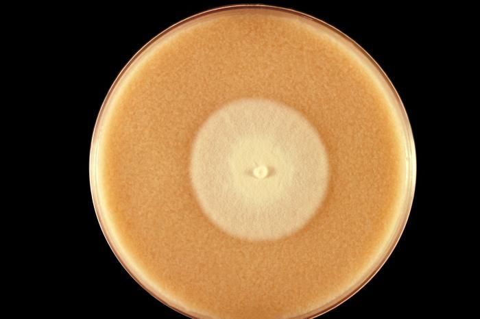
Photographed from the front, this image depicted a Petri dish containing cereal agar, upon which a Microsporum persicolor fungal colony had been cultured. As was the case here, the colonies can be white, or depending upon the Microsporum sp., may run the gamut, sporting a yellow, beige or cinnamon color, and display a flat, or glabrous, woolly or powdery texture. Taxonomically, M. persicolor is a member of the phylum Ascomycota. See PHIL 10905 for a reverse view of this colony, i.e., viewed from behind.Created: 1973
-
Viewed from the back, i.e., reverse, this image depicted a Petri dish containing Sabouraud's (SAB) dextrose agar, upon which a Microsporum persicolor fungal colony had been cultured. As seen in this reverse view, the colonial coloration can be yellow, or may even be a red-brown. From the front, as depicted in PHIL 10906, the colonies can be white, or depending upon the Microsporum sp., may run the gamut, sporting a yellow, beige or cinnamon color, and display a flat, or glabrous, woolly or powdery texture.Created: 1973
-
Photographed from the front, this image depicted a Petri dish containing cereal agar, upon which a Microsporum persicolor fungal colony had been cultured. As was the case here, the colonies can be white, or depending upon the Microsporum sp., may run the gamut, sporting a yellow, beige or cinnamon color, and display a flat, or glabrous, woolly or powdery texture. Taxonomically, M. persicolor is a member of the phylum Ascomycota. See PHIL 10903 for a reverse view of this colony, i.e., viewed from behind.Created: 1973
-
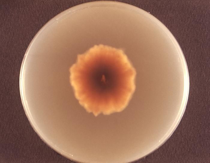
Viewed from the back, i.e., reverse, this image depicted a Petri dish containing Sabouraud's (SAB) dextrose agar, upon which a Microsporum persicolor fungal colony had been cultured. As seen in this reverse view, the colonial coloration can be yellow, or may even be a red-brown. From the front, as depicted in PHIL 10904, the colonies can be white, or depending upon the Microsporum sp., may run the gamut, sporting a yellow, beige or cinnamon color, and display a flat, or glabrous, woolly or powdery texture.Created: 1973
-
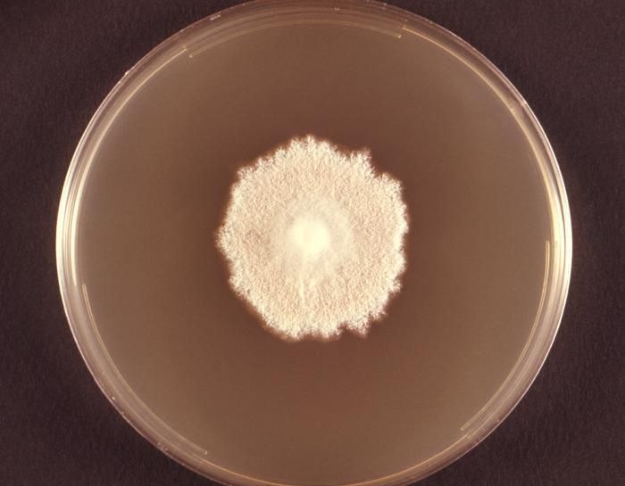
Photographed from the front, this image depicted a Petri dish containing cereal agar, upon which a Microsporum persicolor fungal colony had been cultured. As was the case here, the colonies can be white, or depending upon the Microsporum sp., may run the gamut, sporting a yellow, beige or cinnamon color, and display a flat, or glabrous, woolly or powdery texture. Taxonomically, M. persicolor is a member of the phylum Ascomycota.Created: 1973
-
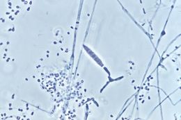
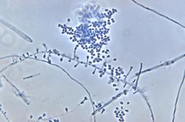
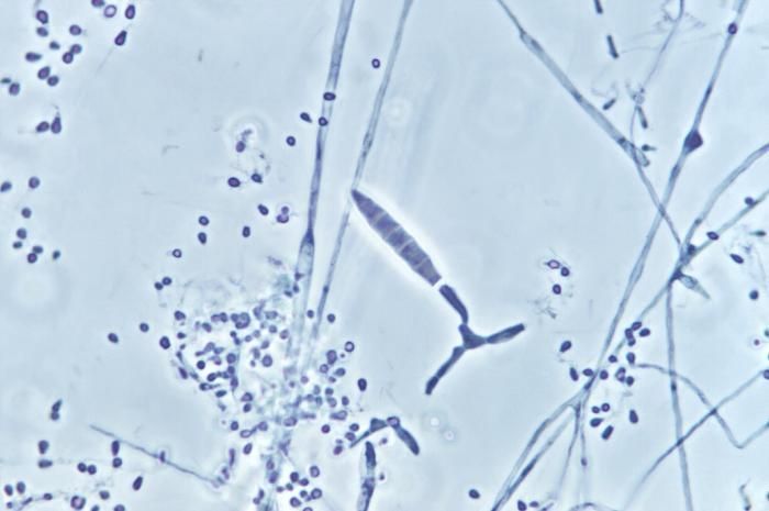
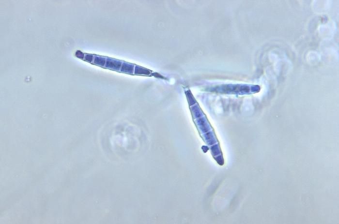
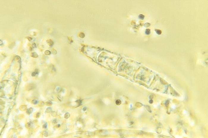
Magnified 500X, this photomicrograph revealed some of the ultrastructural morphology exhibited by the fungal organism, Microsporum persicolor. In this particular image details seen in both a centrally-located macroconidium, and numerous, single-celled microconidia are revealed. Both of these structure types are the asexual spores that originate from the filamentous conidiophore, and are also known as mitospores, for they are born out of the process of mitosis, and are therefore, haploid when they reach maturity. Unlike the single-celled microconidia, the M. persicolor macroconidia are composed of multiple, attached microconidia, separtated by cell walls, and configured in a cigar-shaped chain.Created: 1973
-
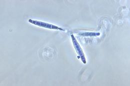
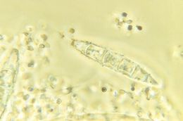
Magnified 500X, this photomicrograph revealed some of the ultrastructural morphology exhibited by the fungal organism, Microsporum persicolor. This particular image highlights details seen in three macroconidia, which are the asexual spores that originate from the filamentous conidiophore. These macroconidia are also known as mitospores, for they are born out of the process of mitosis, and are therefore, haploid when they reach maturity. Unlike the single-celled microconidia, the M. persicolor macroconidia are composed of multiple, attached microconidia, separtated by cell walls, and configured in a cigar-shaped chain.Created: 1973
-
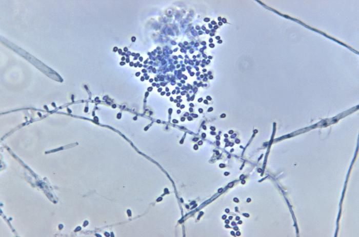
Magnified 500X, this photomicrograph revealed some of the ultrastructural morphology exhibited by the fungal organism, Microsporum persicolor. Of particular note are the numerous microconidia configured both in clusters, and as singular units. These microconidia are the asexual spores and originate from the filamentous conidiophore. These conidia are also known as mitospores, for they are born out of the process of mitosis, and are therefore, haploid when they reach maturity.Created: 1973
-
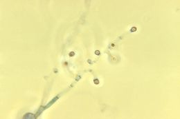
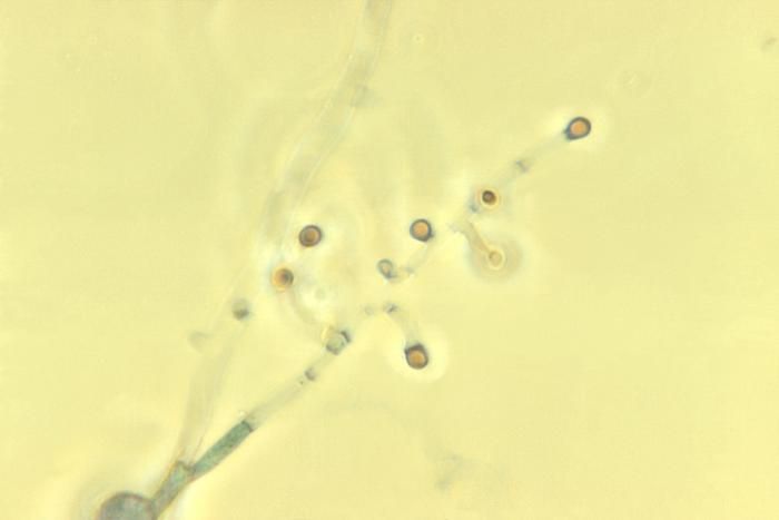
This photomicrograph depicted a number of Microsporum persicolor fungal microconidia. Under this relatively-high magnification of 1500X, the ultrastructural morphology exhibited by these spores is revealed.Created: 1973
-
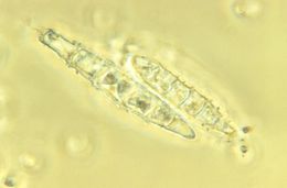
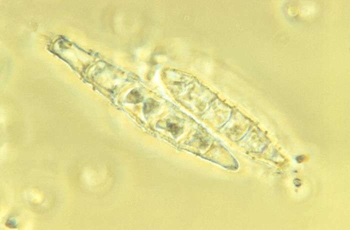
Characterized as echinulate, or spiny, this photomicrograph depicted a number of Microsporum persicolor fungal macroconidia. Under this relatively-high magnification of 1125X, the ultrastructural morphology exhibited by these elongated, roughened spore clusters can be observed.Created: 1973
-
Characterized as echinulate, or spiny, this photomicrograph depicted a number of Microsporum persicolor fungal macroconidia. Under this relatively-high magnification of 1125X, the ultrastructural morphology exhibited by these elongated, roughened spore clusters can be observed.Created: 1973
-
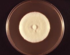
This is the top view of a Sabourauds dextrose plate culture growing the fungus Microsporum persicolor.Created: 1973
-
This is the bottom or reverse view of a cereal agar plate culture growing the fungus Microsporum persicolor.Created: 1973
-
This is a top view of a cereal agar plate culture growing the fungus Microsporum persicolor.Created: 1973
-
This is a top view of a Sabourauds dextrose agar plate culture growing Microsporum persicolor.Created: 1973
-
This is the bottom or reverse view of a Sabourauds dextrose plate culture growing the fungus Microsporum persicolor.Created: 1973
-
This is a top view of a cereal agar plate culture of a Microsporum persicolor fungal colony.Created: 1973
-
This is a top view of a Sabourauds dextrose agar plate culture of a Microsporum persicolor fungal colony.Created: 1973
-
This is the bottom or reverse view of a Sabourauds dextrose plate culture growing the fungus Microsporum persicolor.Created: 1973
-
This is a top view of a cereal agar plate culture growing the fungus Microsporum persicolor.Created: 1973
-
This is a top view of a Sabourauds dextrose agar plate culture growing Microsporum persicolor.Created: 1973
-
This is the bottom, or reverse view of a Sabourauds dextrose agar plate culture of Microsporum persicolor.Created: 1973
-
This is a top view of a cereal agar plate culture growing a colony of Microsporum persicolor fungus.Created: 1973