Plate 18
Descrição:
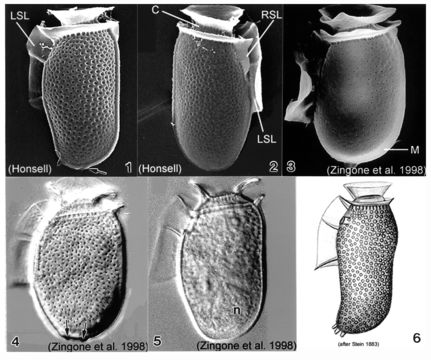
Plate 18. Dinophysis sacculus. Figs. 1-3. SEM: lateral view. Fig. 1. Cell oblong with rounded posterior. Hypotheca long, margins undulate. Thecal surface coarsely areolated. Short left sulcal list (LSL). Cingulum with two well developed lists. Small blunt posterior projections (arrow). Fig. 2. Cingulum lined with pores. Right sulcal list (RSL) visible. Fig. 3. Smooth thecal surface with pores. Metacytic zone (M) devoid of pores. Figs. 4-5. LM: lateral view. Fig. 4. Hypotheca sack-like with deep thecal pores. Posterior end with two blunt projections (arrows). Fig. 5. Large posterior nucleus (n). Fig. 6. Line drawing: morphotype from Stein (1883).
Incluído nas seguintes páginas:
- Life
- Cellular
- Eukaryota
- SAR (Stramenopiles, Alveolates, Rhizaria)
- Alveolata
- Dinophyceae
- Dinophysiales
- Dinophysiaceae
- Dinophysis
- Dinophysis sacculus
- Dinoflagellata
Esta imagem não aparece em nenhuma coleção.
Informação de origem
- licença
- cc-publicdomain
- citação bibliográfica
- Faust, Maria A. and Rose A. Gulledge. Identifying Harmful Marine Dinoflagellates. Smithsonian Contributions from the United States National Herbarium, volume 42: 1-144 (including 48 plates, 1 figure and 1 table).
- original
- arquivo de mídia original
- visite a fonte
- site do parceiro
- NMNH Marine Dinoflagellates
- ID