Imagem de Giardia
Descrição:
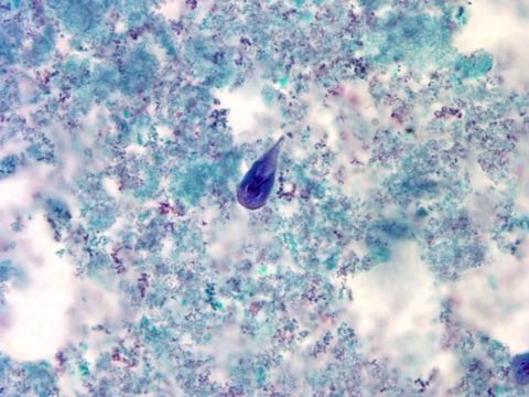
At a magnification of 1000X, this trichrome-stained photomicrograph revealed the morphologic characteristics of a blue-stained Giardia intestinalis protozoan trophozoite (center). In the small intestine, the protozoan cysts release trophozoites, with each cyst producing two trophozoites. Trophozoites multiply by longitudinal binary fission, remaining in the lumen of the proximal small bowel where they can be free, or attached to the mucosa by a ventral sucking disk. As the trophozoites mature, they are simultaneously migrating towards the colon, whereupon, they once again become thick-walled cysts, and are in this way, passed in the hosts stool into the environment. As cysts, these protozoan parasites can survive for many months until they are accidentally ingested by another unfortunate host.
Created:
Incluído nas seguintes páginas:
- Life
- Cellular
- Eukaryota
- Excavates
- Metamonada
- Fornicata
- Diplomonadida
- Hexamitidae
- Giardia
- Giardia lamblia
Esta imagem não aparece em nenhuma coleção.
Informação de origem
- licença
- cc-publicdomain
- fornecedor
- Public Health Image Library
- original
- arquivo de mídia original
- visite a fonte
- site do parceiro
- Public Health Image Library
- ID