Imagem de Hymenomonas roseola Stein 1878
Descrição:
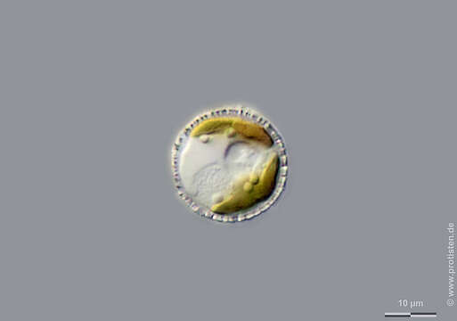
Sampling date 05/2017. Scale bars indicate 10 µm.
Four images. Cells in immotile stage.
First couple:Top view showing calcareous scales, followed by a cross-section showing chloroplast and storage globules.Second couple:Top view showing calcareous scales, followed by an image with a cross-section showing parietal chloroplasts, storage globules, a contractile vacuole and the nucleus (granulated area side by side to the cv).Please click on < or > on the image edges or on the dots at the bottom edge of the images to browse through the slides!
Place name: Pond in the forest of Altenholz-Stift (Schleswig-Holstein, Germany)
Latitude: 54.384913 Longitude: 10.125691
Microscope Zeiss Axioplan, camera Olympus OM-D M5 MKII. DOF images.
© Wolfgang Bettighofer,
images under Creative Commons License V 3.0 (CC BY-NC-SA).
For permission to use of (high resolution) images please contact postmaster@protisten.de.
For further information about the image, please click here: Link to protisten.de page
Incluído nas seguintes páginas:
- Life
- Cellular
- Eukaryota
- Haptista
- Haptophyta
- Prymnesiophyceae
- Coccolithales
- Hymenomonadaceae
- Hymenomonas
- Hymenomonas roseola
Esta imagem não aparece em nenhuma coleção.
Informação de origem
- licença
- cc-by-nc-sa-3.0
- direitos autorais
- Wolfgang Bettighofer
- criador
- Wolfgang Bettighofer [email]
- original
- arquivo de mídia original
- visite a fonte
- site do parceiro
- protisten.de
- ID