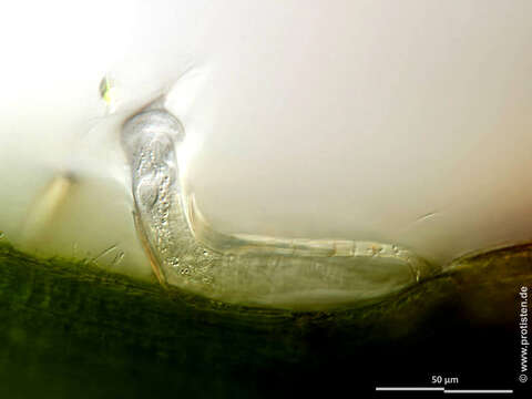
Imagem de Platycola longicollis Kent 1882
Descrição:
Scale bars indicate 50 µm.
Seven images, six of them in a slide changer.
First (in the slide changer):Complete representation of the trophont and its lorica.Second:Optical cross-section showing the elliptical shaped vestibulum.Third and fourth:Optical cross-section. The sectional plane is placed in such a way that the striation on the cell body is partially shown (arrows and insets in fourth image). Objective 40x/1.1 water immersion.Fifth and sixth:Optical cross-section through the cell and the lorica showing several parts of the worm-shaped macronucleus traversing almost the entire cell (arrows and insets in sixth image). The brightly glowing dots in the cell body are mitochondria.Please click on < or > on the image edges or on the dots at the bottom edge of the images to browse through the slides!
Place name: Tropical freshwater aquarium
Latitude: 54.3018013 Longitude: 10.07120132
Microscope Zeiss Axioplan, camera Olympus OM-D M5 MKII. DOF images.
© Wolfgang Bettighofer,
images under Creative Commons License V 3.0 (CC BY-NC-SA).
For permission to use of (high resolution) images please contact postmaster@protisten.de.
For further information about the image, please click here: Link to protisten.de page
Incluído nas seguintes páginas:
- Life
- Cellular
- Eukaryota
- SAR (Stramenopiles, Alveolates, Rhizaria)
- Alveolata
- Ciliophora
- Intramacronucleata
- Oligohymenophorea
- Peritrichia
- Sessilida
- Vaginicolidae
- Platycola
- Platycola longicollis
Esta imagem não aparece em nenhuma coleção.
Informação de origem
- licença
- cc-by-nc-sa-3.0
- direitos autorais
- Wolfgang Bettighofer
- criador
- Wolfgang Bettighofer [email]
- original
- arquivo de mídia original
- visite a fonte
- site do parceiro
- protisten.de
- ID