in vivo, dorsal pellicular detail
Descrição:
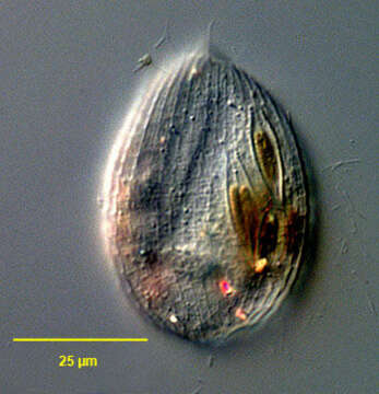
Portrait (dorsal view) of the oligohymenophorean ciliate, Lembadion lucens (Maskell, 1887) Kahl, 1931 showing detail of the pellicle. The cell outline is oval. The ventral surface is concave and the dorsum convex. The very large scoop-like peristome occupies most of the ventral surface. The cytostome is at the posterior end of the peristome. There is a small undulating membrane on the right margin of the peristome. A large sheet-like adoral membranelle arises from the left margin of the peristome. There are 25-35 evenly spaced longitudinal somatic kineties. The posterior 2/3 of the pellicle of L. lucens has an areolate pattern divided into small roughly rectangular depressions (similar to the pattern of the entire pellicle of L. bullinum). The dikinetids of the somatic kineties occupy the center of the rectangles. The anterior 1/3 of the pellicle has a longitudinal striate pattern (similar to the pattern of the entire pellicle of L. magnum). The somatic dikinetids lie in the center of these striae. Ingested diatoms and highly refractile crystals are visible in the cytoplasm. Collected from a freshwater pond near Boise, Idaho July 2004. DIC .
Incluído nas seguintes páginas:
- Life
- Cellular
- Eukaryota
- SAR (Stramenopiles, Alveolates, Rhizaria)
- Alveolata
- Ciliophora
- Intramacronucleata
- Oligohymenophorea
- Peniculida
- Lembadionidae
- Lembadion
- Lembadion lucens
Esta imagem não aparece em nenhuma coleção.
Informação de origem
- licença
- cc-by-nc
- autor
- William Bourland
- fornecedor
- micro*scope
- original
- arquivo de mídia original
- visite a fonte
- site do parceiro
- micro*scope
- ID