Dorsal view, in vivo
Descrição:
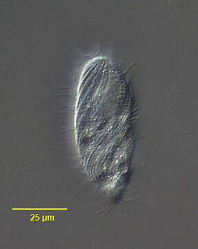
Portrait (dorsal view) of the trichostomatid ciliate, Spirozona caudata (Kahl,1926). The cell is elongate and rounded anteriorly and tapers posteriorly to a narrow truncate cone. The cell is ellipsoid in cross section. The cytostome is located in the anterior ¼. Its right margin is curved and the left relatively straight. There are several oral polykinetids on the left and an undulating membrane on the right. The somatic ciliature is distinctive with a wide swath of closely spaced kineties spiraling from the right anterior to the posterior midline. A single spiral kinety of more densely packed kinetosomes bearing longer cilia originates to the right of the cytostome and spirals around the right side to the dorsum of the cell. There are 3 postoral kineties and one short paraoral kinetid on the left margin of the cytostome. More widely spaced kineties with less densely packed kinetids originate to the left of the cytostome and follow a less spiral course to the posterior end ventrally. The narrow truncate cone of the posterior end bears a circular row of kinetids. There is a small unciliated anterior apical area or "frontal plate". The spherical macronucleus and micronucleus are located in the anterior half. The single contractile vacuole is located at the posterior end. Collected from sapropelic bottom sediments from standing freshwater near Boise, Idaho November, 2004. DIC.
Incluído nas seguintes páginas:
- Life
- Cellular
- Eukaryota (Eucariontes)
- SAR (Stramenopiles, Alveolates, Rhizaria)
- Alveolata
- Ciliophora
- Intramacronucleata
- Oligohymenophorea
- Hymenostomatida
- Spirozonidae
- Spirozona
- Spirozona caudata
Esta imagem não aparece em nenhuma coleção.
Informação de origem
- licença
- cc-by-nc
- autor
- William Bourland
- fornecedor
- micro*scope
- original
- arquivo de mídia original
- visite a fonte
- site do parceiro
- micro*scope
- ID