PhycRes-pre12392-fig-0001a-m-Vampirovibrio-chlorellavorus
Descrição:
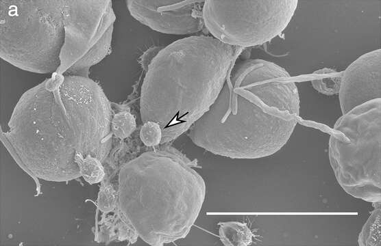
Description: English: Scanning Electron Micrograph of Chlorella sorokiniana and attached Vampirovibrio chlorellavorus cells. Image of a sample collected from Arizona test site at 10 000 × magnification with V. chlorellavorus indicated by the white arrow.Scale bar is displayed in white representing 5.0 μm. Date: 17 July 2019. Source: Fig. 1a at https://onlinelibrary.wiley.com/doi/10.1111/pre.12392 Vampirovibrio chlorellavorus draft genome sequence, annotation, and preliminary characterization of pathogenicity determinants Phycological Research Vol. 68, No. 1 p. 23-29, doi:10.1111/pre.12392 . Author: Blake T. Hovde, Seth A. Steichen, Shawn R. Starkenburg, Judith K. Brown. Other versions:.
Incluído nas seguintes páginas:
- Life
- Cellular
- Eukaryota
- Archaeplastida
- Chloroplastida
- Chlorophyta
- Trebouxiophyceae
- Chlorellales
- Chlorellaceae
- Chlorella (Clorela)
Esta imagem não aparece em nenhuma coleção.
Informação de origem
- licença
- cc-by-sa-3.0
- direitos autorais
- Blake T. Hovde, Seth A. Steichen, Shawn R. Starkenburg, Judith K. Brown
- criador
- Blake T. Hovde, Seth A. Steichen, Shawn R. Starkenburg, Judith K. Brown
- fonte
- Fig. 1a at https://onlinelibrary.wiley.com/doi/10.1111/pre.12392 Vampirovibrio chlorellavorus draft genome sequence, annotation, and preliminary characterization of pathogenicity determinants Phycological Research Vol. 68, No. 1 p. 23-29, doi:10.1111/pre.12392
- original
- arquivo de mídia original
- visite a fonte
- site do parceiro
- Wikimedia Commons
- ID