black and white portrait
Descrição:
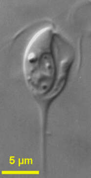
Chilomastix (kai-low-ma-sticks) cuspidata (Larsen and Patterson, 1990) Bernard et al., 1997. Cells are drop-shaped with a long posterior spike and about 20 - 32 microns long (including the spike) with a groove extending from the apex to the posterior end of the untapered part of the cell. The cells have 4 flagella inserting subapically and are directed anterior laterally, one is shorter than the cell and the other three are about the cell length. The short flagellum beats and lies within the ventral groove. The nucleus is situated subapically. Food vacuoles occur throughout the cell. The cells move slowly by swimming while rotating and may attach to the substrate by the tip of the spike.
Incluído nas seguintes páginas:
- Life
- Cellular
- Eukaryota (Eucariontes)
- Excavates
- Metamonada
- Fornicata
- Retortamonadidae
- Chilomastix
- Chilomastix cuspidata
Esta imagem não aparece em nenhuma coleção.
Informação de origem
- licença
- cc-by-nc
- autor
- Won Je Lee
- fornecedor
- micro*scope
- original
- arquivo de mídia original
- visite a fonte
- site do parceiro
- micro*scope
- ID