portrait
Descrição:
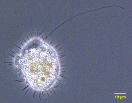
Portrait of Mastigina setosa a flagellated pelobiont. The ameboid monopodial cells have radiating "setae" which are 6-10 micron long filaments. There is a single, long, lazily beating flagellum, which is closely associated with the nucleus (seen at the 1 o clock position adjacent to the cell membrane in this image). Electron microscopy shows the base of the flagellum connected to the nucleus by a cone of microtubules. The flagellum has a variable arrangement of microtubules. The nucleus contains a large nucleolus. Ingested algae are seen in food vacuoles. There is vigorous cytoplasmic streaming resulting in ameboid locomotion. The flagellum appears to contribute little to motility. Mastigina and the other pelobionts lack mitochondria and dictyosomes. Obvious ameboid locomotion and cytoplasmic streaming may help differentiate Mastigina from Mastigamoeba, a similar pelobiont. From slow-moving organically enriched freshwater runoff stream near Boise, Idaho. Phase contrast.
Incluído nas seguintes páginas:
- Life
- Cellular
- Eukaryota (Eucariontes)
- Amoebozoa
- Evosea
- Mastigamoebidae
- Mastigina
- Mastigina setosa
- Archamoebea
Esta imagem não aparece em nenhuma coleção.
Informação de origem
- licença
- cc-by-nc
- autor
- William Bourland
- fornecedor
- micro*scope
- original
- arquivo de mídia original
- visite a fonte
- site do parceiro
- micro*scope
- ID