Multi-layer image
Descrição:
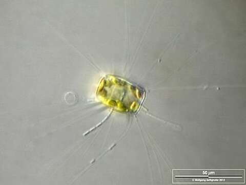
Optical tranversal section, showing the nucleus. The thin, barely visible floating extensions are made of chitin. Furthermore, filamentous bacteria colonies are attached. Scale bar indicates 50 µm.
Sample from the Lake Constance (vicinity of Bodman). The image was built up using several photomicrographic frames with manual stacking technique. Images were taken using Zeiss Universal with Olympus C7070 CCD camera.
Image under Creative Commons License V 3.0 (CC BY-NC-SA).
Incluído nas seguintes páginas:
- Life
- Cellular
- Eukaryota (Eucariontes)
- SAR (Stramenopiles, Alveolates, Rhizaria)
- Stramenopiles
- Oomycota
- Ochrophyta
- Diatomista
- Bacillariophyta
- Coscinodiscophyceae
- Thalassiosirophycidae
- Thalassiosirales
- Stephanodiscaceae
- Kuetzing
Esta imagem não aparece em nenhuma coleção.
Informação de origem
- licença
- cc-by-nc
- fornecedor
- micro*scope
- original
- arquivo de mídia original
- visite a fonte
- site do parceiro
- micro*scope
- ID