Imagem de Mcvaughia
Descrição:
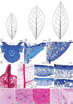
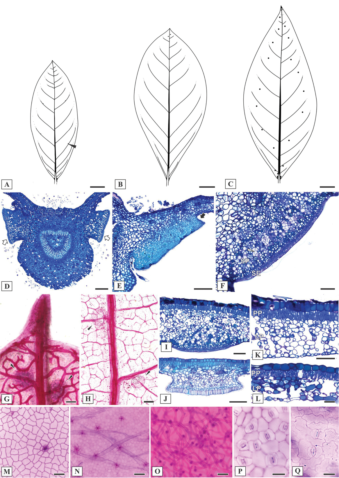
Figure 3. Leaf morphoanatomy of Mcvaughia species. A patterns of leaf glands distribution on the abaxial leaf surface of M.bahianaB patterns of leaf glands distribution on the abaxial leaf surface of M.piauhiensisC patterns of leaf glands distribution on the abaxial leaf surface of M.sergipanaD transverse section of leaf base showing the basilaminar pair of stalked glands (white arrows) E basilaminar leaf gland with a stalk (black arrow) in M.piauhiensisF basilaminar gland in M.sergipana showing a sessile position (SE= anatomical arrangement with secretory epidermis, SP= vascularized secretory parenchyma) G–H laminar glands on the apex of cleared leaves of M.sergipana and M.bahiana respectively, note the apical tooth (G) I sessile laminar glands in M.sergipanaJ stalked laminar gland in M.piauhiensisK–L transverse sections of the leaf blade; mesophyll with uniserial palisade-like parenchyma and spongy parenchyma composed by several or few layers in M.sergipana and M.bahiana, respectively; note the idioblast with druse crystals at the mesophyll (white arrow) and the stomata distribution at the abaxial leaf surface (black arrow) M–N adaxial epidermis surface of M.piauhiensis and M.sergipana, showing scars of malpighiaceous trichomes O abaxial epidermis surface of trichomes abundance in M.bahianaP–Q outline of the anticlinal epidermal cell walls: straight in M.sergipana (P) and sinuous in M.bahiana (Q). Laminar scale bars: 1 cm (A–C), 100 μm (D, F–K, N–O), 150 μm (E), 50 μm (L–M, P–Q).
Incluído nas seguintes páginas:
- Life
- Cellular
- Eukaryota
- Archaeplastida
- Chloroplastida
- Spermatophytes (Spermatophyta)
- Angiosperms
- Eudicots
- Superrosids
- Rosids
- Malpighiales
- Malpighiaceae
- Mcvaughia
- NO NAME!
Esta imagem não aparece em nenhuma coleção.
Informação de origem
- licença
- cc-by-3.0
- direitos autorais
- Rafael F. Almeida, Isabel R. Guesdon, Marcelo R. Pace, Renata M.S. Meira
- citação bibliográfica
- Almeida R, Guesdon I, Pace M, Meira R (2019) Taxonomic revision of Mcvaughia W.R.Anderson (Malpighiaceae): notes on vegetative and reproductive anatomy and the description of a new species PhytoKeys (117): 45–72
- original
- arquivo de mídia original
- visite a fonte
- site do parceiro
- Phytokeys
- ID