dcsimg

Description of Micromonas

provided by BioPedia
Extremely small flagellate with single emergent flagellum, single parietal chloroplast and single pyrenoid. Eyespot absent. Cells naked. The axoneme-containing part of the flagellum very short but the central pair of microtubules extends into a long thin whip. This is usually visible only in the electron microscope. Healthy cells swim quickly with the short stub-like part of the flagellum directed backwards. One species.
license
cc-by-nc
author
biopedia
provider
BioPedia
original
visit source
partner site
BioPedia

Micromonas

provided by wikipedia EN

Micromonas is a genus of green algae in the family Mamiellaceae.[1][2]

Before the characterization in 2016 of a second species, Micromonas commoda, Micromonas pusilla was considered to be the only species in the genus,[3][4] which led to a disproportionate amount of research discussing a single species within the genus. It is suggested to be the dominant photosynthetic picoeukaryote in some marine ecosystems.[5] Unlike many marine algae, it is distributed widely in both warm and cold waters.[6] It is a strong swimmer and exhibits a phototaxic response.[6]

Micromonas pusilla is divided into 3 to 5 different clades despite their similarity in morphologies and habitats.[7][8] Varying ratios of clades contribute to the M. pusilla population throughout the marine ecosystem leading to the hypothesis of clades arising based on niche occupation and susceptibility to virus infection.[8]

Discovery

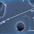
Micromonas pusilla is considered the first picoplankton studied, when it was discovered and named Chromulina pusilla in the 1950s by R. Butcher.[9] Later, electron micrographs by the English scientists, Irene Manton and Mary Park, in the 1960s provided further details on M. pusilla.[9]

Cell morphology and structure

Micromonas is a group of small unicellular pear-shaped micro-algae that do not have a cell wall.[10][11][12] Just like other members in the class, they have a single mitochondrion and a single chloroplast,[12] which covers almost half of the cell.[12][13] They are able to swim due to the presence of a scale-less flagellum.[12][13][3] The axonemal structure of the flagellum for this genus is different in that the peripheral microtubules do not extend up to the termination of the central pair of microtubules, allowing a visible investigation of the motion of the central pair.[11][14][15] In Micromonas, the central pair constantly rotates in an anti-clockwise direction, despite the motion of other components of the flagellum.[11][14]

While the cell size, shape and the location of insertion of the flagellum into the cell are similar among strains and genetic clades, the variation in respective hair point length results in different lengths of the flagella within the genus.[3]

Antibiotic

The antibiotic susceptibility was determined using a single strain of M. pusilla with the purpose to produce axenic cultures to be used in studies and experiments.[16] The strain of M.pusilla was tested with a range of antibiotics to determine the possible effects of the particular antibiotic.[16]

Resistance:[16] benzylpenicillin, gentamicin, kanamycin, neomycin, streptomycin

Sensitive:[16] chloramphenicol, polymyxin B

For M. pusilla, sensitivity towards an antibiotic is likely defined by the impairment of growth, rather than a lethal effect, when subjected to bactericidal levels of that particular antibiotic.[16] The susceptibility of other strains of M. pusilla towards this set of antibiotics should be the same.[16]

Genetics

Evolutionary history

Micromonas diverged early on from the lineage that led to all modern terrestrial plants. Individual species have very similar 18S ribosomal RNA gene sequences, a comparison often used to determine microscopic speciation, however, only 90% of different genes are shared between all Micromonas species. This presence or absence of complete genes, compared with only small changes of sequence to specific genes, suggests that Micromonas is the result of intensive horizontal gene transfer.[10]

Strain Isolation

The original Micromonas reference genome was created from a strain, RCC299, first isolated in 1998 from an Equatorial Pacific sample. This strain has been continuously cultured for two decades and is available from the Roscoff Culture Collection. In 2005, a monoclonal culture of the strain was isolated. The axenic strain is available from the Center for Culture of Marine Phytoplankton, under the name CCMP2709. Currently, a separate stain that was isolated from temperate coastal waters is being sequenced.[10]

Genome Structure

The entire Micromonas sp. genome was first shotgun sequenced in 2014. Micromonas has about 19Mb, however this varies slightly between species and strain. This is made up of 17 chromosomes and contains 59% GC content.[17] Based on [open reading frames] the genome codes for about 10000 proteins and 70 functional RNAs.[18]

Cellular mechanisms

Cell growth and division

Micromonas reproduces asexually through fission.[11] It has been observed that M. pusilla shows variability in optical characteristics, for example cell size and light scattering, throughout the day.[19] There is an increase in these measurements during the period with light, followed by a decrease during period without light.[19][20] This coincides with the findings that proteomic profiles change over the diel cycle, with an increase in expression of proteins related to cell proliferation, lipid and cell membrane restructuring in the dark when cells start dividing and become smaller.[20] However, the expression levels of genes and proteins can still vary within the same metabolic pathway.[20] It has also been suggested that the structure of 3’ UTR may play a role in the regulatory system.[20]

Light-harvesting system

Micromonas species still share the same collection of photosynthetic pigments as the members of the class Mamiellophyceae,[3] which includes the common pigments chlorophyll a and chlorophyll b,[21] as well as prasinoxanthin (xanthophyll K), the first algal carotenoid being assigned with a structure that has a γ-end group.[22] It has been discovered that most of its xanthophylls are in the oxidized state and show similarities to ones possessed by other important marine planktons like diatoms, golden and brown algae, and dinoflagellates.[23] In addition, there is another pigment called Chl cCS-170 can be found in some strains of Micromonas and Ostreococcus living in deeper part of the ocean, which may indicate a potential adaptation for organisms that reside under low light intensity.[3]

The light-harvesting complexes of Micromonas are distinguishable from other green algae in terms of pigment composition and stability under unfavorable conditions.[21] It has been showed that these proteins use three different pigments for light harvesting, and they are resistant to high temperature and the presence of detergent.

Peptidoglycan biosynthesis

Even though the chloroplasts, which are suggested to be originated from Cyanobacteria via endosymbiosis,[24] from Micromonas do not have a surrounding peptidoglycan layer, the peptidoglycan biosynthesis pathway is found to be complete in M. pusilla and partial in M. commoda, with the presence of some relevant enzymes only.[12] While the role of this pathway for Micromonas is still under investigation, this observation shows a lineage for different species of Micromonas along with glaucophyte algae which still have their chloroplasts covered with peptidoglycan.[12]

Ecological significance

Micromonas make up a significant amount of picoplanktonic biomass and productivity in both oceanic and coastal regions.[5] The abundance of Micromonas has increased over the past decade. Evidence shows these spikes in numbers are induced through climate change, which has been felt more drastically in the Arctic.[12] In past years it was thought that green algal species were solely photosynthetic only to discover that was not the case.[25] Micromonas, as well as the Prasinophytes, have taken on mixotrophic lifestyles and have huge impacts on prokaryotic populations within the Arctic.[25] Due to the large consumption of prokaryotes by Micromonas, studies are suggesting photosynthetic picoeukaryotes will soon dominate the primary productivity and bacterivory of Arctic systems.[25] Laboratory studies have shown that species within a single genus have altered their mixotrophic strategies to their distinct environments.[25] These environments can differ through light intensity, nutrient availability and size of prey which all have shown clade-specific alterations to maximize efficiencies.[25]

Viral infection

Viruses are important in the balance of marine ecosystem by regulating the composition of microbial communities, but their behaviors can be affected by several factors including temperature, mode of infection and host conditions.[26][27] There is an increasing number of Micromonas-infecting virus being discovered and studied.

Micromonas pusilla virus

There are currently 45 viral strains identified that coexist with M. pusilla populations.[8] Virus infectivity is dependent on the host strain, light availability and virus adsorption.[28]

Per day average of death due to virus lysis is estimated to be about 2 to 10% of the M. pusilla population.[28]

  • Micromonas pusilla reovirus (MpRV): The first isolation of a reovirus that infects protist.[29] This virus is found to be bigger than other members of the family.[30]

Micromonas polaris virus

It is the first phycodnavirus being isolated from polar ocean waters.[31] It can infect M. polaris, which is the polar ecotype of Micromonas that has adapted to waters with low temperatures.[31]

Evidence suggests that the increase in temperature due to climate change may shift the clonal composition of both the virus and host.[31]

Metabolic engineering

With the growing population in the world, there is an increased demand for wild fishes and algae for their source of polyunsaturated fatty acids (PUFA), which is required for growth and development, as well as the maintenance of health in humans. Recent research is investigating an alternative mechanism for production of PUFA by using acyl-CoA Δ6-desaturase, an enzyme present in M. pusilla, with plants. The M. pusilla strain of acyl-CoA Δ6-desaturase is highly effective in the polyunsaturated fatty acid synthesis pathway due to its strong binding preference for omega-3 substrates in land plants.[32]

References

  1. ^ See the NCBI webpage on Micromonas. Data extracted from the NCBI taxonomy resources, National Center for Biotechnology Information, retrieved 2007-03-19
  2. ^ Micromonas Manton & Parke, 1960, non Borrel, 1902, World Register of Marine Species, accessed March 6, 2010
  3. ^ a b c d e Simon, Nathalie; Foulon, Elodie; Grulois, Daphné; Six, Christophe; Desdevises, Yves; Latimier, Marie; Gall, Florence Le; Tragin, Margot; Houdan, Aude (2017). "Revision of the Genus Micromonas Manton et Parke (Chlorophyta, Mamiellophyceae), of the Type Species M. pusilla (Butcher) Manton & Parke and of the Species M. commoda van Baren, Bachy and Worden and Description of Two New Species Based on the Genetic and Phenotypic Characterization of Cultured Isolates" (PDF). Protist. 168 (5): 612–635. doi:10.1016/j.protis.2017.09.002. PMID 29028580.
  4. ^ Borowitzka, Michael A.; Beardall, John; Raven, John A. (2016-03-21). The physiology of microalgae. Borowitzka, Michael A.,, Beardall, John,, Raven, John A. Cham. ISBN 9783319249452. OCLC 945445086.
  5. ^ a b Not, F; Latasa, M; Marie, D; Cariou, T; Vaulot, D; Simon, N (Jul 2004), "A Single Species, Micromonas pusilla (Prasinophyceae), Dominates the Eukaryotic Picoplankton in the Western English Channel", Applied and Environmental Microbiology, 70 (7): 4064–72, Bibcode:2004ApEnM..70.4064N, doi:10.1128/AEM.70.7.4064-4072.2004, ISSN 0099-2240, PMC 444783, PMID 15240284
  6. ^ a b Genomes of Two Strains of Micromonas Algae Show Surprising Diversity Archived 2011-07-07 at the Wayback Machine, Alternative Energy Newswire, April 10, 2009
  7. ^ Foulon, Elodie; Not, Fabrice; Jalabert, Fabienne; Cariou, Thierry; Massana, Ramon; Simon, Nathalie (1 September 2008). "Ecological niche partitioning in the picoplanktonic green alga Micromonas pusilla: evidence from environmental surveys using phylogenetic probes". Environmental Microbiology. 10 (9): 2433–2443. doi:10.1111/j.1462-2920.2008.01673.x. PMID 18537812.
  8. ^ a b c Baudoux, A.-C.; Lebredonchel, H.; Dehmer, H.; Latimier, M.; Edern, R.; Rigaut-Jalabert, F.; Ge, P.; Guillou, L.; Foulon, E.; Bozec, Y.; Cariou, T.; Desdevises, Y.; Derelle, E.; Grimsley, N.; Moreau, H.; Simon, N. (1 October 2015). "Interplay between the genetic clades of Micromonas and their viruses in the Western English Channel" (PDF). Environmental Microbiology Reports. 7 (5): 765–773. doi:10.1111/1758-2229.12309. PMID 26081716.
  9. ^ a b Vaulot, Daniel; Eikrem, Wenche; Viprey, Manon; Moreau, Hervé (1 August 2008). "The diversity of small eukaryotic phytoplankton (≤3 μm) in marine ecosystems". FEMS Microbiology Reviews. 32 (5): 795–820. doi:10.1111/j.1574-6976.2008.00121.x. PMID 18564290.
  10. ^ a b c Worden, Alexandra Z.; Lee, Jae-Hyeok; Mock, Thomas; Rouzé, Pierre; Simmons, Melinda P.; Aerts, Andrea L.; Allen, Andrew E.; Cuvelier, Marie L.; Derelle, Evelyne (2009-04-10). "Green Evolution and Dynamic Adaptations Revealed by Genomes of the Marine Picoeukaryotes Micromonas". Science. 324 (5924): 268–272. Bibcode:2009Sci...324..268W. doi:10.1126/science.1167222. ISSN 0036-8075. PMID 19359590. S2CID 206516961.
  11. ^ a b c d Bell, Peter R. (2000). Green plants : their origin and diversity. Hemsley, Alan R. (2nd ed.). Cambridge, UK: Cambridge University Press. ISBN 978-0-521-64109-8. OCLC 56124600.
  12. ^ a b c d e f g van Baren, Marijke J.; Bachy, Charles; Reistetter, Emily Nahas; Purvine, Samuel O.; Grimwood, Jane; Sudek, Sebastian; Yu, Hang; Poirier, Camille; Deerinck, Thomas J. (2016-03-31). "Evidence-based green algal genomics reveals marine diversity and ancestral characteristics of land plants". BMC Genomics. 17: 267. doi:10.1186/s12864-016-2585-6. ISSN 1471-2164. PMC 4815162. PMID 27029936.
  13. ^ a b Advances in marine biology. Volume 60. Lesser, Michael. Amsterdam: Elsevier Academic Press. 2011. ISBN 978-0-12-385529-9. OCLC 761362752.{{cite book}}: CS1 maint: others (link)
  14. ^ a b Omoto, Charlotte K.; Witman, George B. (1981-04-23). "Functionally significant central-pair rotation in a primitive eukaryotic flagellum". Nature. 290 (5808): 708–710. Bibcode:1981Natur.290..708O. doi:10.1038/290708a0. ISSN 1476-4687. PMID 7219555. S2CID 4354444.
  15. ^ Vaulot, Daniel; Eikrem, Wenche; Viprey, Manon; Moreau, Hervé (2008-08-01). "The diversity of small eukaryotic phytoplankton (≤3 μm) in marine ecosystems". FEMS Microbiology Reviews. 32 (5): 795–820. doi:10.1111/j.1574-6976.2008.00121.x. ISSN 0168-6445. PMID 18564290.
  16. ^ a b c d e f Cottrell, Matthew T.; Suttle, Curtis A. (1 June 1993). "Production of Axenic Cultures of Micromonas Pusilla (Prasinophyceae) Using Antibiotic 1". Journal of Phycology. 29 (3): 385–387. doi:10.1111/j.0022-3646.1993.00385.x. S2CID 85052488.
  17. ^ NCBI Micromonas Genome Repository https://www.ncbi.nlm.nih.gov/genome/2297
  18. ^ JPI KEGG genome, micromonas comoda http://www.genome.jp/kegg-bin/show_organism?org=mis
  19. ^ a b DuRand, Michele D.; Green, Rebecca E.; Sosik, Heidi M.; Olson, Robert J. (2002-12-01). "Diel Variations in Optical Properties of Micromonas Pusilla (prasinophyceae)1". Journal of Phycology. 38 (6): 1132–1142. doi:10.1046/j.1529-8817.2002.02008.x. ISSN 1529-8817. S2CID 28859691.
  20. ^ a b c d Waltman, Peter H.; Guo, Jian; Reistetter, Emily Nahas; Purvine, Samuel; Ansong, Charles K.; Baren, Marijke J. van; Wong, Chee-Hong; Wei, Chia-Lin; Smith, Richard D. (2016-07-19). "Identifying Aspects of the Post-Transcriptional Program Governing the Proteome of the Green Alga Micromonas pusilla". PLOS ONE. 11 (7): e0155839. Bibcode:2016PLoSO..1155839W. doi:10.1371/journal.pone.0155839. ISSN 1932-6203. PMC 4951065. PMID 27434306.
  21. ^ a b Wilhelm, C.; Lenartz-Weiler, I.; Wiedemann, I.; Wild, A. (1986). "The light-harvesting system of a Micromonas species (Prasinophyceae): the combination of three different chlorophyll species in one single chlorophyll–protein complex". Phycologia. 25 (3): 304–312. doi:10.2216/i0031-8884-25-3-304.1.
  22. ^ Foss, Per; Guillard, Robert R.L.; Liaaen-Jensen, Synnøve (1984). "Prasinoxanthin—a chemosystematic marker for algae". Phytochemistry. 23 (8): 1629–1633. doi:10.1016/s0031-9422(00)83455-x.
  23. ^ Ricketts, T.R. (1966). "The carotenoids of the phytoflagellate, Micromonas pusilla". Phytochemistry. 5 (4): 571–580. doi:10.1016/s0031-9422(00)83635-3.
  24. ^ Machida, Mariko; Takechi, Katsuaki; Sato, Hiroshi; Chung, Sung Jin; Kuroiwa, Haruko; Takio, Susumu; Seki, Motoaki; Shinozaki, Kazuo; Fujita, Tomomichi (2006-04-25). "Genes for the peptidoglycan synthesis pathway are essential for chloroplast division in moss". Proceedings of the National Academy of Sciences. 103 (17): 6753–6758. Bibcode:2006PNAS..103.6753M. doi:10.1073/pnas.0510693103. PMC 1458953. PMID 16618924.
  25. ^ a b c d e McKie-Krisberg, Zaid M; Sanders, Robert W (October 2014). "Phagotrophy by the picoeukaryotic green alga Micromonas: implications for Arctic Oceans". The ISME Journal. 8 (10): 1953–1961. doi:10.1038/ismej.2014.16. PMC 4184008. PMID 24553471.
  26. ^ Demory, David; Arsenieff, Laure; Simon, Nathalie; Six, Christophe; Rigaut-Jalabert, Fabienne; Marie, Dominique; Ge, Pei; Bigeard, Estelle; Jacquet, Stéphan (March 2017). "Temperature is a key factor in Micromonas–virus interactions". The ISME Journal. 11 (3): 601–612. doi:10.1038/ismej.2016.160. ISSN 1751-7370. PMC 5322312. PMID 28085157.
  27. ^ Maat, Douwe S.; Bleijswijk, Van; L, Judith D.; Witte, Harry J.; Brussaard, Corina P. D. (2016-09-01). "Virus production in phosphorus-limited Micromonas pusilla stimulated by a supply of naturally low concentrations of different phosphorus sources, far into the lytic cycle". FEMS Microbiology Ecology. 92 (9): fiw136. doi:10.1093/femsec/fiw136. ISSN 0168-6496. PMID 27316561.
  28. ^ a b Cottrell, Matthew T.; Suttle, Curtis A. (1 June 1995). "Dynamics of lytic virus infecting the photosynthetic marine picoflagellate Micromonas pusilla". Limnology and Oceanography. 40 (4): 730–739. Bibcode:1995LimOc..40..730C. doi:10.4319/lo.1995.40.4.0730.
  29. ^ Brussaard, C.P.D; Noordeloos, A.A.M; Sandaa, R.-A; Heldal, M; Bratbak, G (2004). "Discovery of a dsRNA virus infecting the marine photosynthetic protist Micromonas pusilla". Virology. 319 (2): 280–291. doi:10.1016/j.virol.2003.10.033. PMID 14980488.
  30. ^ Attoui, H; Jaafar, Fm; Belhouchet, M; De, Micco, P; De, Lamballerie, X; Brussaard, Cp (May 2006), "Micromonas pusilla reovirus: a new member of the family Reoviridae assigned to a novel proposed genus (Mimoreovirus)" (Free full text), The Journal of General Virology, 87 (Pt 5): 1375–83, doi:10.1099/vir.0.81584-0, ISSN 0022-1317, PMID 16603541{{citation}}: CS1 maint: multiple names: authors list (link)
  31. ^ a b c Maat, Douwe S.; Biggs, Tristan; Evans, Claire; van Bleijswijk, Judith D. L.; van der Wel, Nicole N.; Dutilh, Bas E.; Brussaard, Corina P. D. (2017-06-02). "Characterization and Temperature Dependence of Arctic Micromonas polaris Viruses". Viruses. 9 (6): 134. doi:10.3390/v9060134. PMC 5490811. PMID 28574420.
  32. ^ Petrie, James R.; Shrestha, Pushkar; Mansour, Maged P.; Nichols, Peter D.; Liu, Qing; Singh, Surinder P. (1 May 2010). "Metabolic engineering of omega-3 long-chain polyunsaturated fatty acids in plants using an acyl-CoA Δ6-desaturase with ω3-preference from the marine microalga Micromonas pusilla". Metabolic Engineering. 12 (3): 233–240. doi:10.1016/j.ymben.2009.12.001. PMID 20004733.

license
cc-by-sa-3.0
copyright
Wikipedia authors and editors
original
visit source
partner site
wikipedia EN

Micromonas: Brief Summary

provided by wikipedia EN

Micromonas is a genus of green algae in the family Mamiellaceae.

Before the characterization in 2016 of a second species, Micromonas commoda, Micromonas pusilla was considered to be the only species in the genus, which led to a disproportionate amount of research discussing a single species within the genus. It is suggested to be the dominant photosynthetic picoeukaryote in some marine ecosystems. Unlike many marine algae, it is distributed widely in both warm and cold waters. It is a strong swimmer and exhibits a phototaxic response.

Micromonas pusilla is divided into 3 to 5 different clades despite their similarity in morphologies and habitats. Varying ratios of clades contribute to the M. pusilla population throughout the marine ecosystem leading to the hypothesis of clades arising based on niche occupation and susceptibility to virus infection.

license
cc-by-sa-3.0
copyright
Wikipedia authors and editors
original
visit source
partner site
wikipedia EN

Classification

provided by World Register of Marine Species
Classified under family Mamiellaceae in aphia
license
cc-by-4.0
copyright
WoRMS Editorial Board
contributor
Simon Claus [email]