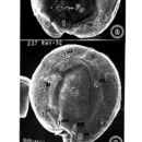
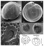
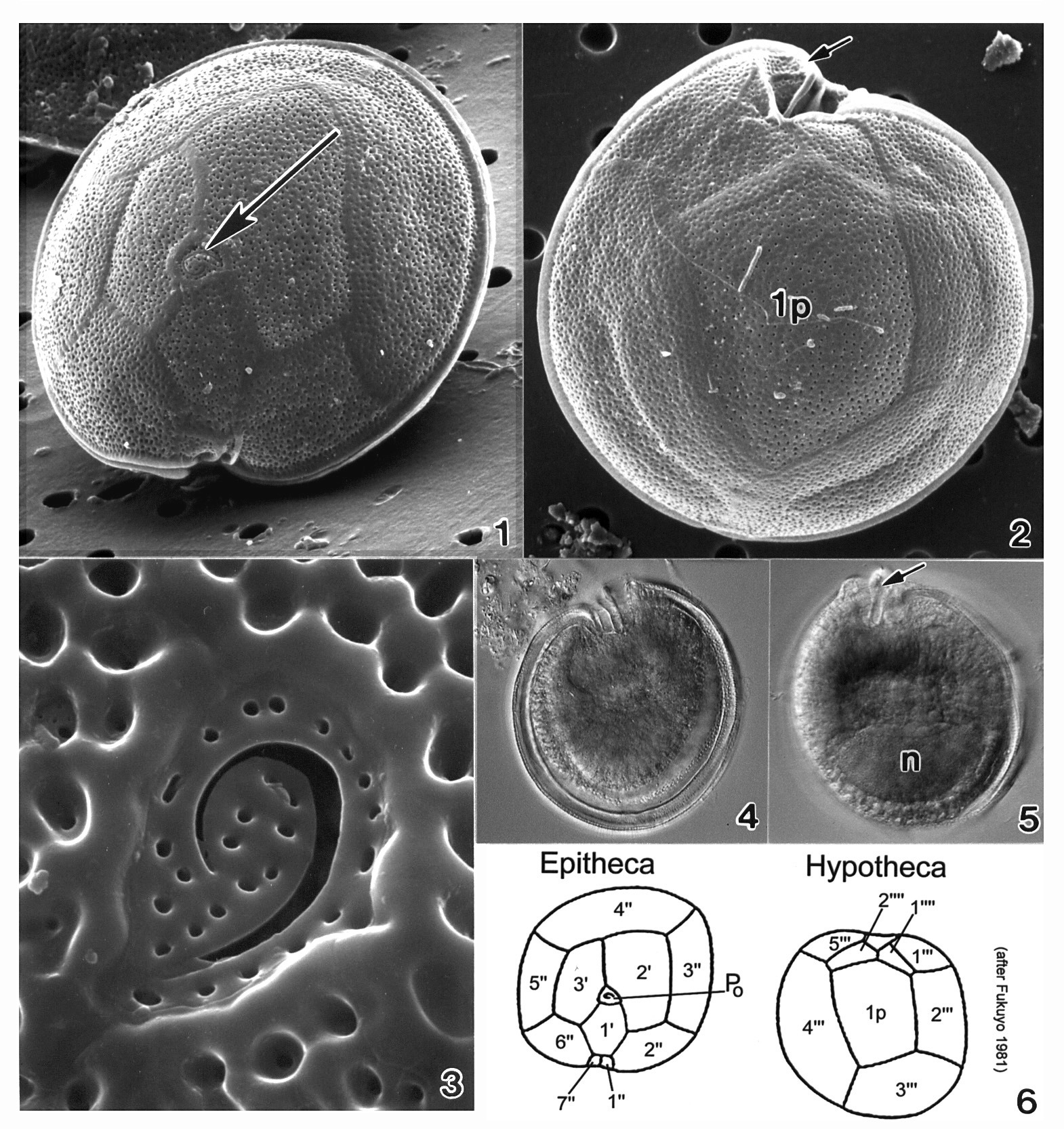
Plate 20. Gambierdiscus toxicus. Figs. 1-3. SEM. Fig. 1. Epitheca: cell round to ellipsoid; anterior-posteriorly compressed. Cell surface smooth with small scattered pores. Apical pore complex located at the apex (arrow). Fig. 2. Hypotheca: 1p plate large and pentagonal. Sulcal region deeply excavated (arrow). Fig. 3. Apical pore plate with characteristic fishhook shaped apical pore. Fig. 4. LM. Epitheca: cingulum and sulcal region in focus. Fig. 5. LM. Hypotheca: sulcal ridge (arrow); large nucleus (n). Fig. 6. Line drawing: thecal plate arrangement.