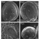
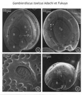
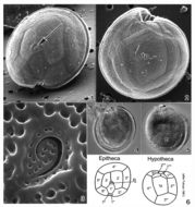
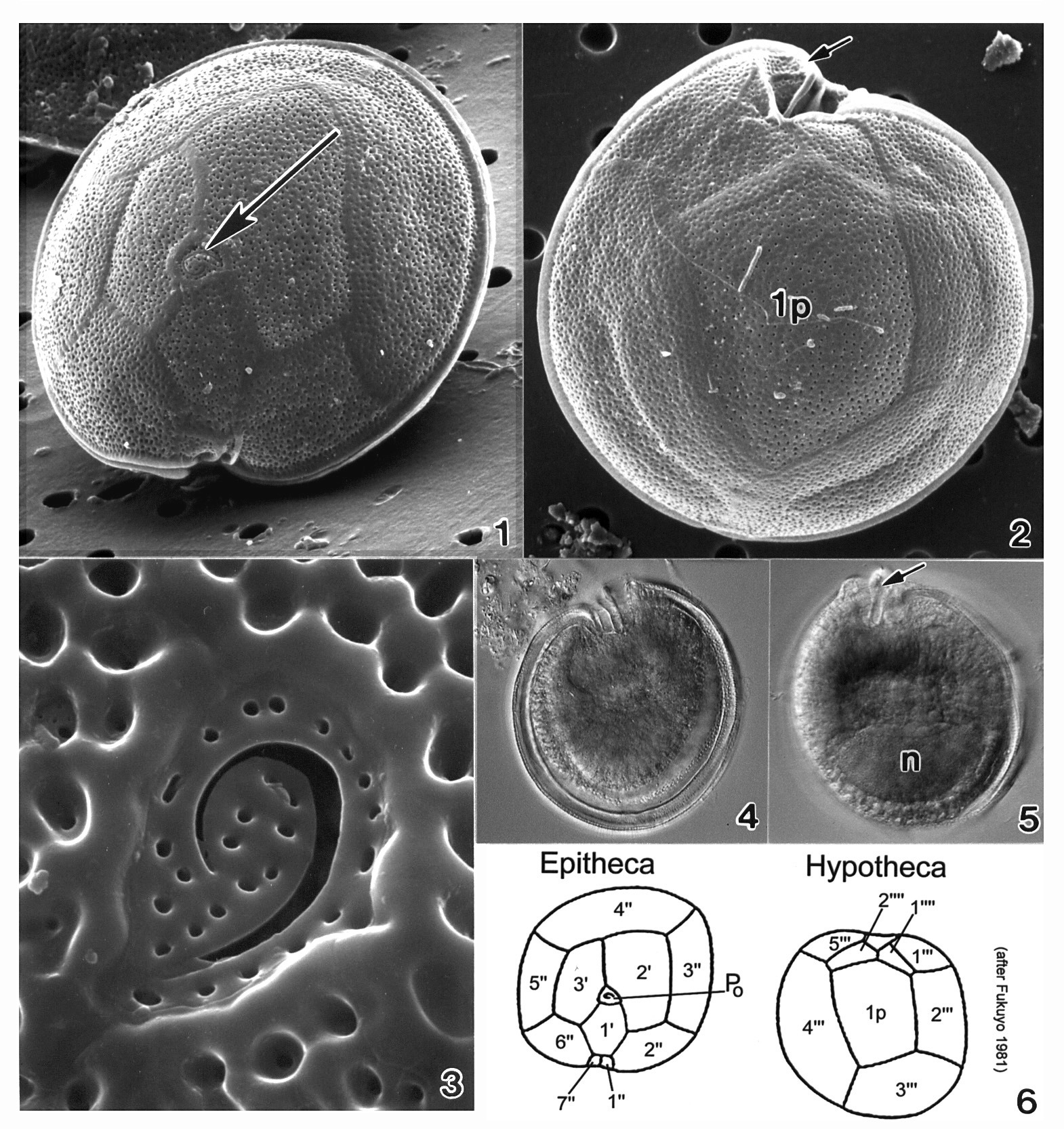
Figs. 1-4. Scanning electron micrographs illustrate the surface morphology of Gambierdiscus toxicus (GTT-91). Fig. 1. In epithecal view. The cell shape is round, and the apical pore plate (Po) oriented ventrally. Fig. 2. In hypothecal view. Posterior intercalary plate (Ip) broad and pentagonal, centrally located, occupying about 1/3 of cell's width. Fig.3. Po plate ellipsoid with a fish-hook-shaped apical pore surrounded by rows of 28 evenly distributed pores. Fig. 4. Cell in central view, shape compressed and bordered by a cingular list. The cell surface is smooth with small scattered pores.
NOTE: This is the apotype of the genus Gambierdiscus. It is an important toxic species. I would like to add this species to the dinoflagellate ‘Types’ since the SEM plate of G. toxicus is the only record. Adachi and Fukuyo (1979) described G. toxicus sp. nov. only in line drawing to illustrate the morphology of plates.
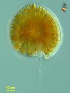
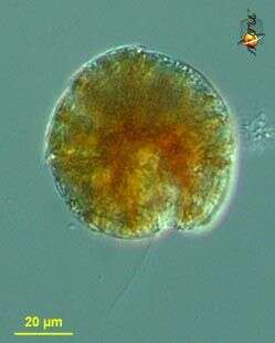
Gambierdiscus (gam-beer-disk-us) toxicus, a toxic dinoflagellate with chloroplasts. With two flagella, but only one (the trailing flagellum) visible here. Small lobes of the plastid extend to the surface of the cell. Armoured, with fairly thick thecal plates. Flattened and this view is more or less polar. Mostly associated with sediments of warmer waters. Differential interference microscopy. data on this strain.
Gambierdiscus (gam-beer-disk-us) toxicus, a toxic dinoflagellate with chloroplasts. With two flagella, but only one (the trailing flagellum) visible here. Armoured, with fairly thick thecal plates. Flattened and this view is more or less polar. Mostly from warmer waters. Differential interference microscopy. data on this strain.
Gambierdiscus (gam-beer-disk-us) toxicus, a toxic dinoflagellate with chloroplasts (not shown in this image). Armoured, with fairly thick thecal plates. This image shows the plates and a large apical pore. Mostly from sediments of warmer waters. Differential interference microscopy. data on this strain.