-
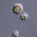
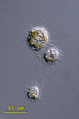
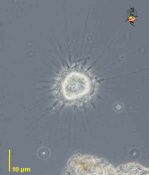
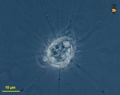
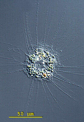
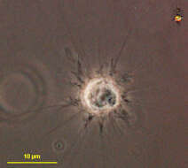
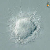
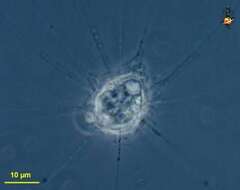
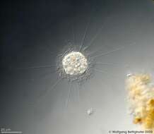
Raphidiophrys (raff-fid-ee-off-riss) ambigua is a solitary species in this genus of centrohelid heliozoa. It forms three kinds of spindle shaped spicules that accumulate around the pseudopodia. The first form are spindle shaped and pointed at both ends, the second form are medium sized, broadly spindle shaped and rounded at the ends, and the final form are very small (5 - 6 mm long) and elliptically shaped. The spicules are embedded in a gelatinous envelope of the cell. Cytoplasm without symbiotic algae but often coloured greenish, brownish or yellowish by food vacuoles. Three closely arranged specimen of Raphidiophrys ambigua, possibly shortly after cell division. Note the yellow brownish food vacuoles that are characteristic of this species. From a pond near Konstanz, Germany.Differential interference contrast.
-
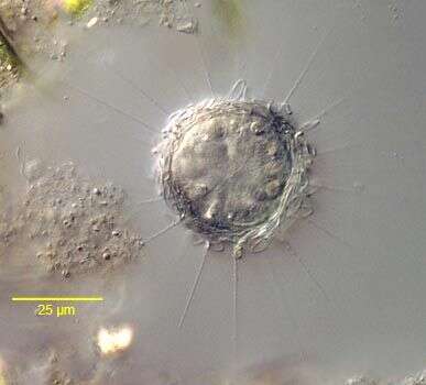
Detail of the siliceous scales of the centroheliozoan, Polyplacocystis symmetrica (Penard, 1904) Mikrjukov, 1996. An axopodium with a bead-like extrusome is visible below the scale to the viewer's left. Collected from a freshwater aquaculture pond near Boise, Idaho. November 2003. DIC.
-
Single whole plate scale viewed by transmission electron microscopy. The siliceous scales are formed within the cell and then form a loose layer or periplast around the outside of the cell.
-
Portrait of the centroheliozoan, Polyplacocystis symmetrica (Penard, 1904) Mikrjukov, 1996. Collected from a freshwater aquaculture pond near Boise, Idaho. November 2003. DIC.
-
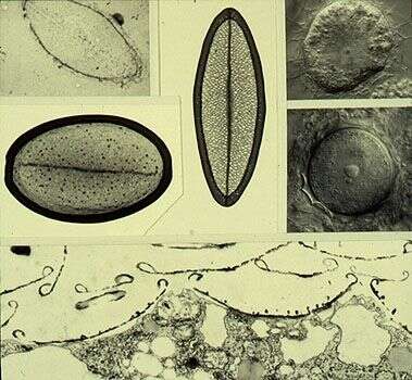
Composite image showing single scales (upper left). Upper left corner treated with hydrofluoric acid to show siliceous nature, vertical scale from trophic cell (Nomarski image upper right corner), larger scale from the cyst (middle right image). The lower image is a transmission electron micrograph of a thin section showing the scales adhering to the outer surface of the cell.
-
Various teratological (malformed) scales. Transmission electron micrographs of whole scales.
-
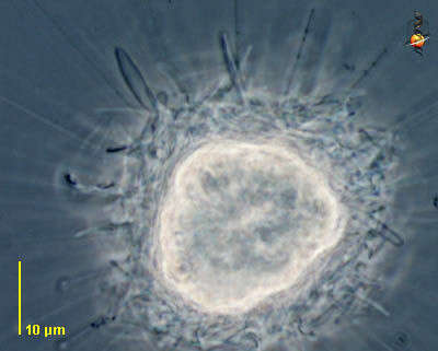
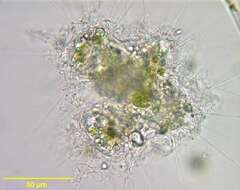
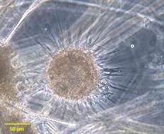
Portrait of centrohelid heliozoan with thick wavy gelatinous mantle incorporating scales. Probably from a genus in the Raphidiophryidae Mikrjukov, 1996, probably Raphidiophrys. This species contains zoochlorellae. From stagnant freshwater pool near Boise, Idaho. I would like to thank Vasilij Zlatogursky of St. Petersburg University for his assistance in identifying this specimen. Brightfield..
-
Portrait of centrohelid heliozoan with thick wavy gelatinous mantle incorporating scales. Probably from a genus in the Raphidiophryidae Mikrjukov, 1996. This species contains zoochlorellae. From stagnant freshwater pool near Boise, Idaho. Phase contrast. I would like to thank Vasilij Zlatogursky of St. Petersburg University for his assistance in identifying this specimen.
-
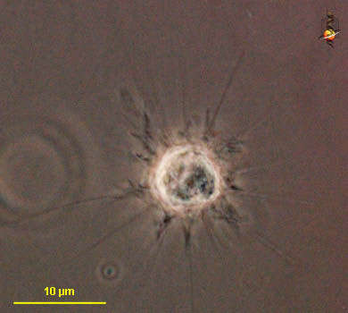
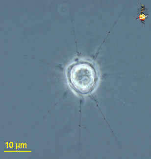
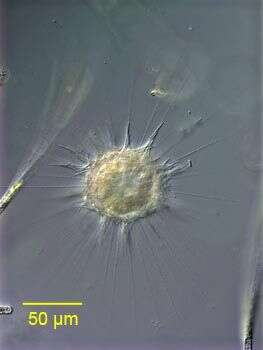
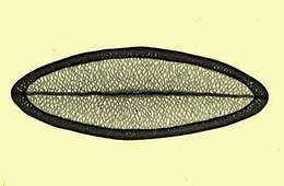
Raphidiophrys (rah-fid-ee-off-riss) is a centrohelid heliozoon. Like other centrohelids, it has thin untapering arms, which have prominent extrusomes. Distinguished from other genera because the cell is enclosed in a layer of loosely adhering boat-shaped scales with thickened margins. Phase contrast.
-
Raphidiophrys (rah-fid-ee-off-riss) is a centrohelid heliozoon. Like other centrohelids, it has thin untapering arms, which have prominent extrusomes. The arms are supported by microtubular axonemes which terminate on a central granule (centroplast) which can just about be seen here. Distinguished from other genera because the cell is enclosed in a layer of loosely adhering boat-shaped scales with thickened margins. Differential interference contrast.
-
Raphidiophrys (rah-fid-ee-off-riss) is a centrohelid heliozoon. Like other centrohelids, it has thin untapering arms, which have prominent extrusomes. Distinguished from other genera because the cell is enclosed in a layer of loosely adhering boat-shaped scales with thickened margins - a good example of which is seen upper left. Phase contrast.
-
Raphidiophrys (rah-fid-ee-off-riss) is a centrohelid heliozoon. Like other centrohelids, it has thin untapering arms, which have prominent extrusomes. Distinguished from other genera because the cell is enclosed in a layer of loosely adhering boat-shaped scales with thickened margins. Phase contrast.
-
-
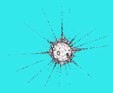
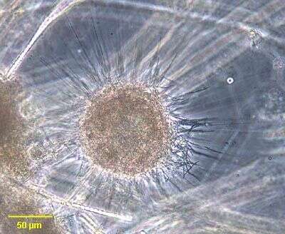
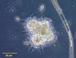
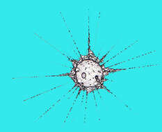
Portrait of Raphidiophrys, a centroheliozoon with siliceous scales but no radiating spicules. Axopodia bearing extrusomes are evident. The abundant scales obscure the details of the cell body in this image. Sometimes multiple individuals cluster. From fresh water pond near Boise, Idaho. Phase contrast.
-
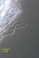
Detail of siliceous tangential scales of Raphidiophrys showing characteristic "canoe" shape. From freshwater pond near Boise, Idaho. Oblique illumination.
-
Raphidiophrys, a centroheliozoon with siliceous scales but no radiating spicules. Axopodia bearing extrusomes are evident. The abundant scales obscure the details of the cell body in this image. Sometimes multiple individuals cluster. From fresh water pond near Boise, Idaho. Phase contrast.
-
-
-
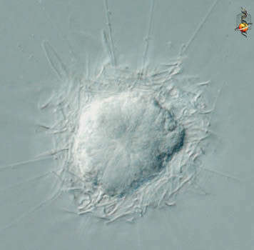
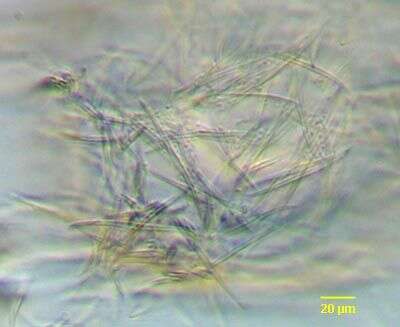
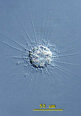
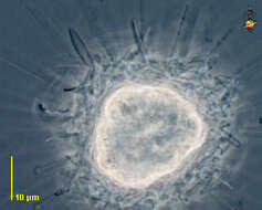
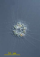
Raphidiophrys elegans can be found either grouped in colonies or as solitary living individuals. In colonies, the disc-shaped spicules form elongate cone-shaped accumulations around the pseudopodia. In solitary living individuals the spicules are arranged more tangentially. The spherical nucleus is placed eccentrically. Usually with one contractile vacuole. Symbiotic algae are sometimes present. Differential interference contrast.
-
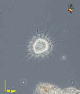
Raphidiophrys elegans can be found either grouped in colonies or as solitary living individuals. In colonies, the disc-shaped spicules form elongate cone-shaped accumulations around the pseudopodia. In solitary living individuals the spicules are arranged more tangentially. The spherical nucleus is placed eccentrically. Usually with one contractile vacuole. Symbiotic algae are sometimes present. A squashed specimen of Raphidiophrys elegans. At about 12:00 the contractile vacuole is visible and the sphaerical nucleus is at 3:00. The spicules are lie tangientially and not cone-shaped around the pseudopodia. From a bog pond near Konstanz, Germany. Differential interference contrast.
-
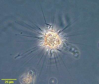
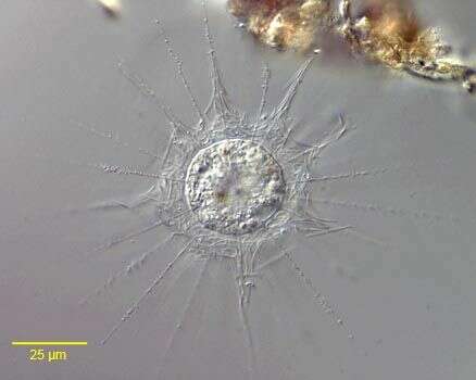
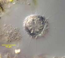
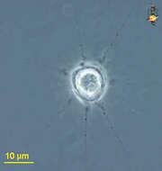
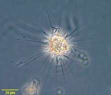
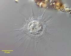
The picture shows the charcteristic layer of mucilage armed with siliceous scales around the cell body, and the fine axopodia with numerous extrusomes. Scale bar indicates 25 µm. Sample from sphagnum pond situated in the northern alpine region of Austria near Salzburg. Images were taken using Zeiss Universal with Olympus C7070 CCD camera.