-
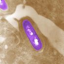
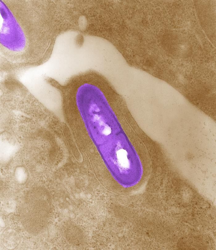
Electron micrograph of a Listeria bacterium in tissue. See PHIL 2286 for a black and white version of this image.Created: 2002
-
This image depicted numbers of Bacillus anthracis bacterial colonies, which had been allowed to grow on sheeps blood agar (SBA) for a 24 hour period. In this particular view youll note the appearance of what is termed a "plaque" (arrowhead), which represents an area where the bacteria had been lysed, or destroyed by the application of a localized amount of gamma phage-containing solution. Highly specific to B. anthracis, these gamma phage viruses, i.e., bacteriophages, attacked the B. anthracis bacteria, subsequently leaving this circular plaque devoid of bacterial organisms. The specificity of these gamma phages to B. anthracis makes this a positive test for the presence of this bacterium.Created: 2009
-
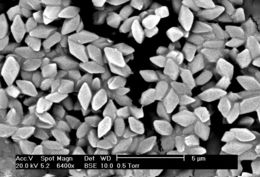
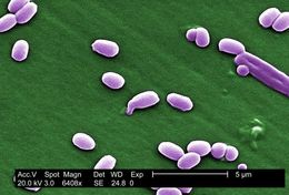
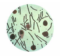
Spores and crystals of Bacillus thuringiensis serovar morrisoni strain T08025 Microscopy by Jim BuckmanFrom
Wikimedia Commons
-
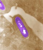
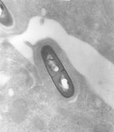
Electron micrograph of a Listeria bacterium in tissue. See PHIL 10828 for a colorized version of this image.Created: 2002
-
This image depicted numbers of Bacillus anthracis bacterial colonies, which had been allowed to grow on sheeps blood agar (SBA) for a 24 hour period. Note the classical appearance exhibited in the colonial morphology including a ground-glass, non-pigmented texture with accompanying comma projections from some of the individual rough-edged colonies. In this particular view, youll note that a tenacity test had been performed using an iinoculating loop, which proved positive for B. anthracis, causing the colony to stand up like beaten egg white.Created: 2009
-
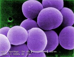
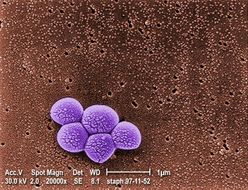
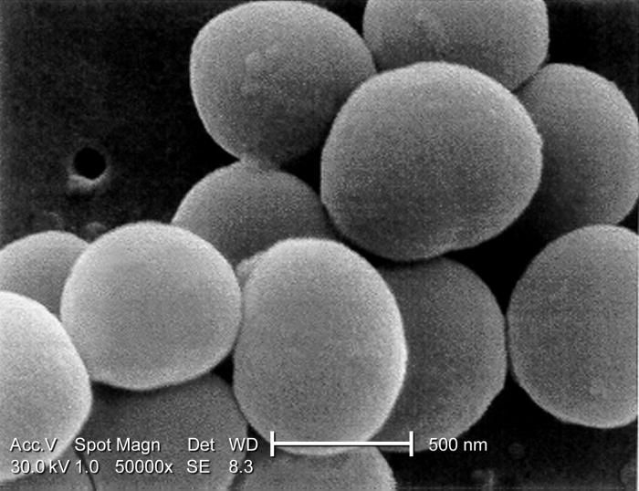
Under a very high magnification of 50,000x, this scanning electron micrograph (SEM) shows a strain of Staphylococcus aureus bacteria taken from a vancomycin intermediate resistant culture (VISA).Under SEM, one can not tell the difference between bacteria that are susceptible, or multidrug resistant, but with transmission electron microscopy (TEM), VISA isolates exhibit a thickening in the cell wall that may attribute to their reduced susceptibility to vancomycin . See PHIL 11158 for a black and white version of this image. VISA and VRSA are specific types of antimicrobial-resistant staph bacteria. While most staph bacteria are susceptible to the antimicrobial agent vancomycin some have developed resistance. VISA and VRSA cannot be successfully treated with vancomycin because these organisms are no longer susceptibile to vancomycin. However, to date, all VISA and VRSA isolates have been susceptible to other Food and Drug Administration (FDA) approved drugs.Created: 2001
-
This image depicted Bacillus anthracis bacterial colonies, which had been allowed to grow on sheeps blood agar (SBA) for a 24 hour period. Note the classical appearance exhibited in the colonial morphology including a ground-glass, non-pigmented texture with accompanying comma projections from some of the individual rough-edged colonies. In this particular view, youll note that a tenacity test had been performed using an iinoculating loop, which proved positive for B. anthracis, causing the colony to stand up like beaten egg white.What is anthrax?Anthrax is an acute infectious disease caused by the spore-forming bacterium Bacillus anthracis. Anthrax most commonly occurs in wild and domestic mammalian species (cattle, sheep, goats, camels, antelopes, and other herbivores), but it can also occur in humans when they are exposed to infected animals or to tissue from infected animals or when anthrax spores are used as a bioterrorist weapon.Created: 2009
-
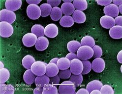
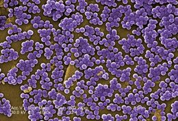
Under a very high magnification of 20,000x, this scanning electron micrograph (SEM) shows a strain of Staphylococcus aureus bacteria taken from a vancomycin intermediate resistant culture (VISA).Under SEM, one can not tell the difference between bacteria that are susceptible, or multidrug resistant, but with transmission electron microscopy (TEM), VISA isolates exhibit a thickening in the cell wall that may attribute to their reduced susceptibility to vancomycin . See PHIL 11156 for a black and white version of this image. VISA and VRSA are specific types of antimicrobial-resistant staph bacteria. While most staph bacteria are susceptible to the antimicrobial agent vancomycin some have developed resistance. VISA and VRSA cannot be successfully treated with vancomycin because these organisms are no longer susceptibile to vancomycin. However, to date, all VISA and VRSA isolates have been susceptible to other Food and Drug Administration (FDA) approved drugs.Created: 2001
-
This image depicted Bacillus anthracis bacterial colonies, which had been allowed to grow on sheeps blood agar (SBA) for a 24 hour period. Note the classical appearance exhibited in the colonial morphology including a ground-glass, non-pigmented texture with accompanying comma projections from some of the individual rough-edged colonies. In this particular view, youll note that a tenacity test had been performed using an iinoculating loop, which proved positive for B. anthracis, causing the colony to stand up like beaten egg white.What is anthrax?Anthrax is an acute infectious disease caused by the spore-forming bacterium Bacillus anthracis. Anthrax most commonly occurs in wild and domestic mammalian species (cattle, sheep, goats, camels, antelopes, and other herbivores), but it can also occur in humans when they are exposed to infected animals or to tissue from infected animals or when anthrax spores are used as a bioterrorist weapon.Created: 2009
-
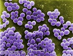
Under a high magnification of 10,000x, this scanning electron micrograph (SEM) shows a strain of Staphylococcus aureus bacteria taken from a vancomycin intermediate resistant culture (VISA).Under SEM, one can not tell the difference between bacteria that are susceptible, or multidrug resistant, but with transmission electron microscopy (TEM), at least with VISA isolates one can see a thickening in the cell wall that may attribute to their reduced susceptibility to vancomycin . See PHIL 11154 for a black and white version of this image.Created: 2001
-
This image depicted numbers of Bacillus anthracis bacterial colonies, which had been allowed to grow on sheeps blood agar (SBA) for a 24 hour period. Note the classical appearance exhibited in the colonial morphology including a ground-glass, non-pigmented texture with accompanying comma projections from some of the individual rough-edged colonies.What is anthrax?Anthrax is an acute infectious disease caused by the spore-forming bacterium Bacillus anthracis. Anthrax most commonly occurs in wild and domestic mammalian species (cattle, sheep, goats, camels, antelopes, and other herbivores), but it can also occur in humans when they are exposed to infected animals or to tissue from infected animals or when anthrax spores are used as a bioterrorist weapon.Created: 2009
-
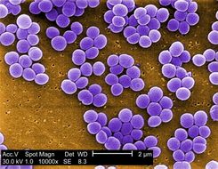
Under a high magnification of 10,000x, this scanning electron micrograph (SEM) shows a strain of Staphylococcus aureus bacteria taken from a vancomycin intermediate resistant culture (VISA).Under SEM, one can not tell the difference between bacteria that are susceptible, or multidrug resistant, but with transmission electron microscopy (TEM), at least with VISA isolates one can see a thickening in the cell wall that may attribute to their reduced susceptibility to vancomycin . See PHIL 6486 for a black and white version of this image.Created: 2001
-
This image depicted numbers of Bacillus anthracis bacterial colonies, which had been allowed to grow on sheeps blood agar (SBA) for a 24 hour period. Note the classical appearance exhibited in the colonial morphology including a ground-glass, non-pigmented texture with accompanying comma projections from some of the individual rough-edged colonies. See PHIL 11748 for a higher magnification of these colonies.What is anthrax?Anthrax is an acute infectious disease caused by the spore-forming bacterium Bacillus anthracis. Anthrax most commonly occurs in wild and domestic mammalian species (cattle, sheep, goats, camels, antelopes, and other herbivores), but it can also occur in humans when they are exposed to infected animals or to tissue from infected animals or when anthrax spores are used as a bioterrorist weapon.Created: 2009
-
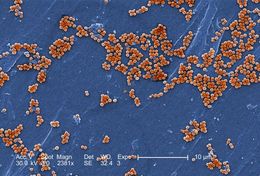
This 2005 scanning electron micrograph (SEM) depicted numerous clumps of methicillin-resistant Staphylococcus aureus bacteria, commonly referred to by the acronym, MRSA; Magnified 2381x. Recently recognized outbreaks, or clusters of MRSA in community settings have been associated with strains that have some unique microbiologic and genetic properties, compared with the traditional hospital-based MRSA strains, which suggests some biologic properties, e.g., virulence factors like toxins, may allow the community strains to spread more easily, or cause more skin disease. A common strain named USA300-0114 has caused many such outbreaks in the United States. See PHIL 7823 for a black and white version of this micrograph.Created: 2005
-
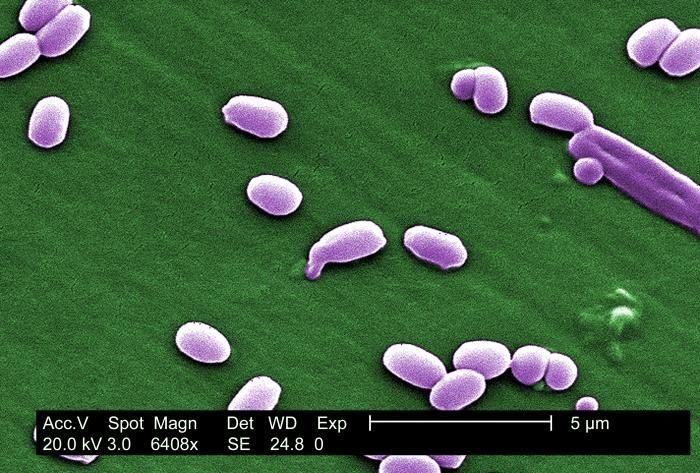
Under a magnification of 6,408X, this scanning electron micrograph (SEM) depicted spores from the Aimes strain of Bacillus anthracis bacteria. See PHIL 10124 for a colorized version of this image.Created: 2002
-
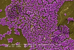
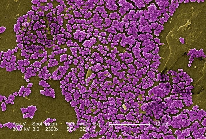
This colorized 2005 scanning electron micrograph (SEM) depicted numerous clumps of methicillin-resistant Staphylococcus aureus bacteria, commonly referred to by the acronym, MRSA; Magnified 2390x. Recently recognized outbreaks, or clusters of MRSA in community settings have been associated with strains that have some unique microbiologic and genetic properties, compared with the traditional hospital-based MRSA strains, which suggests some biologic properties, e.g., virulence factors like toxins, may allow the community strains to spread more easily, or cause more skin disease. A common strain named USA300-0114 has caused many such outbreaks in the United States. Please see PHIL 7822 for a black and white version of this image.Created: 2005
-
Under a very high magnification of 31,207X, this scanning electron micrograph (SEM) depicted spores from the Sterne strain of Bacillus anthracis bacteria. For a black and white version of this image see PHIL 2266.Created: 2002
-
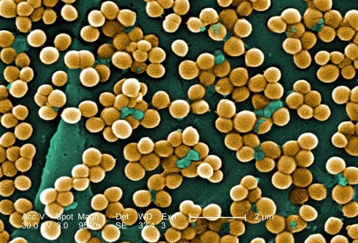
This 2005 scanning electron micrograph (SEM) depicted numerous clumps of methicillin-resistant Staphylococcus aureus bacteria, commonly referred to by the acronym, MRSA; Magnified 9560x.Recently recognized outbreaks, or clusters of MRSA in community settings have been associated with strains that have some unique microbiologic and genetic properties, compared with the traditional hospital-based MRSA strains, which suggests some biologic properties, e.g., virulence factors like toxins, may allow the community strains to spread more easily, or cause more skin disease. A common strain named USA300-0114 has caused many such outbreaks in the United States. See PHIL 7821 for a black and white version of this micrograph.Created: 2005
-
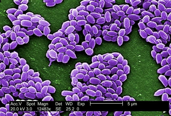
Under a high magnification of 12,483X, this scanning electron micrograph (SEM) depicted spores from the Sterne strain of Bacillus anthracis bacteria. For a black and white version of this image see PHIL 2267.Created: 2002
-
This 2005 colorized scanning electron micrograph (SEM) depicted numerous clumps of methicillin-resistant Staphylococcus aureus bacteria, commonly referred to by the acronym, MRSA; Magnified 4780. Recently recognized outbreaks, or clusters of MRSA in community settings have been associated with strains that have some unique microbiologic and genetic properties, compared with the traditional hospital-based MRSA strains, which suggests some biologic properties, e.g., virulence factors like toxins, may allow the community strains to spread more easily, or cause more skin disease. A common strain named USA300-0114 has caused many such of outbreaks in the United States. See PHIL 7820 for a black and white version of this image.Created: 2005
-
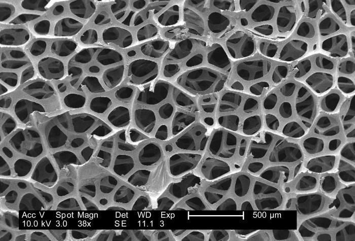
Under a low magnification of 38x, this 2007 scanning electron micrograph (SEM) depicted the fibrous configuration of a dry macrofoam sponge swabs. This swab, as well as three other materials, including polyester (see PHIL 9735), rayon (see PHIL 9734) and cotton (see PHIL 9732, and 9733), were scanned for a CDC study involving their efficiency in recovery of Bacillus anthracis bacterial spores from steel coupons that had been inoculated with a spore suspension of known concentration. See PHIL 9736, 9737, 9738, 9749, 9750, an 9752, for other views of this material. The article discussing the description of this swab material analysis, and the analytical results was published in Emerging Infectious Diseases, Vol. 10, No. 6, June, 2004, and was entitled, Swab Materials and Bacillus anthracis Spore Recovery from Nonporous Surfaces. A link to this article is found below.Created: 2007
-
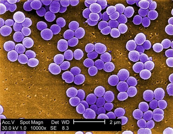
Magnified 20,000X, this colorized scanning electron micrograph (SEM) depicts a grouping of methicillin resistant Staphylococcus aureus (MRSA) bacteria. See PHIL 617 for a black and white view of this image.Created: 1998
-
This illustration depicts Bacillus anthracis taken from the peritoneum using a Hiss capsule stain.Created: 1979
-
Under a very high magnification of 50,000x, this scanning electron micrograph (SEM) shows a strain of Staphylococcus aureus bacteria taken from a vancomycin intermediate resistant culture (VISA).Under SEM, one can not tell the difference between bacteria that are susceptible, or multidrug resistant, but with transmission electron microscopy (TEM), at least with VISA isolates one can see a thickening in the cell wall that may attribute to their reduced susceptibility to vancomycin . See PHIL 11159 for a colorized version of this image. VISA and VRSA are specific types of antimicrobial-resistant staph bacteria. While most staph bacteria are susceptible to the antimicrobial agent vancomycin some have developed resistance. VISA and VRSA cannot be successfully treated with vancomycin because these organisms are no longer susceptibile to vancomycin. However, to date, all VISA and VRSA isolates have been susceptible to other Food and Drug Administration (FDA) approved drugs.Created: 2001


