-
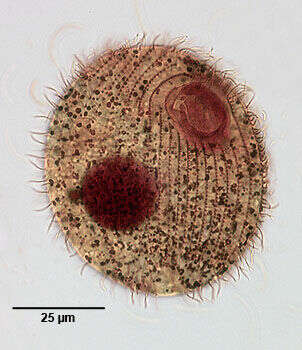
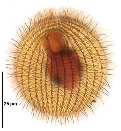
Infraciliature (ventral view) of the hymenostome ciliate, Glaucoma scintillans (Ehrenberg, 1830). Stained by the silver carbonate technic (see Foissner, W. Europ. J. Protistol. 27:313-330;1991). Brightfield
-
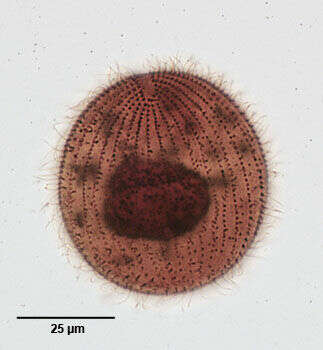
Infraciliature (dorsal view) of the hymenostome ciliate, Glaucoma scintillans (Ehrenberg, 1830). Stained by the silver carbonate technic (see Foissner, W. Europ. J. Protistol. 27:313-330;1991). Brightfield
-
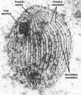
Oral infraciliature of the common hymenostome ciliate, Glaucoma scintillans (EHRENBERG,1830). Stained by the silver carbonate technique (Foissner,W. Europ. J. Protistol.27:313-330;1991).Brightfield.
-
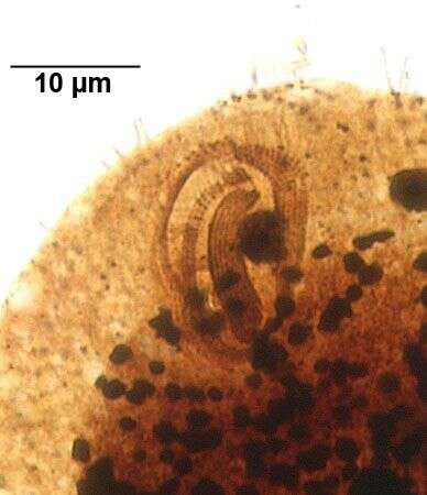
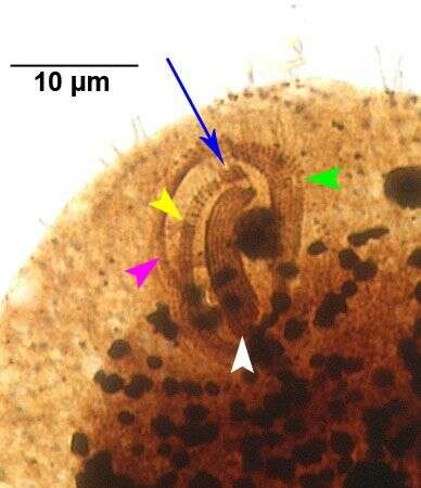
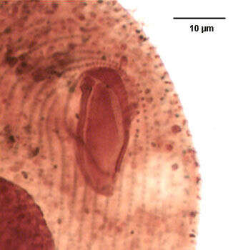
Oral infraciliature of the common hymenostome ciliate, Glaucoma scintillans (EHRENBERG,1830). The three adoral membranelles (M1,M2 and M3) are indicated by the green, white and yellow arrowheads respectively. The "X-body", a rhomboidal group of kinetids just anterior to the end of M2, is indicated by the blue arrow.The undulating membrane on the right margin of the oral aperture is indicated by the pink arrowhead.Stained by the silver carbonate technique (Foissner,W. Europ. J. Protistol.27:313-330;1991).Brightfield.
-
Oral infraciliature of the common hymenostome ciliate, Glaucoma scintillans (EHRENBERG,1830). There are three adoral membranelles (M1,M2 and M3) The "X-body" is a rhomboidal group of kinetids just anterior to the end of M2.Stained by the silver carbonate technique (Foissner,W. Europ. J. Protistol.27:313-330;1991).Brightfield.
-
Infraciliature (ventral view) of the hymenostome ciliate, Glaucoma scintillans (Ehrenberg, 1830). Stained by the silver carbonate technic (see Foissner, W. Europ. J. Protistol. 27:313-330;1991). Brightfield.
-
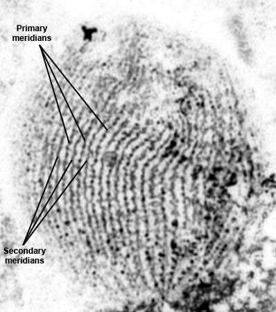
Silverline system of the hymenostome ciliate, Glaucoma scintillans (Ehrenberg, 1830). Stained by the dry silver nitrate technic (see Foissner, W. Europ. J. Protistol. 27:313-330;1991). Brightfield.
-
Silverline system (ventrolateral view) of the hymenostome ciliate, Glaucoma scintillans (Ehrenberg, 1830). Stained by the dry silver nitrate technic (see Foissner, W. Europ. J. Protistol. 27:313-330;1991). Brightfield.
-
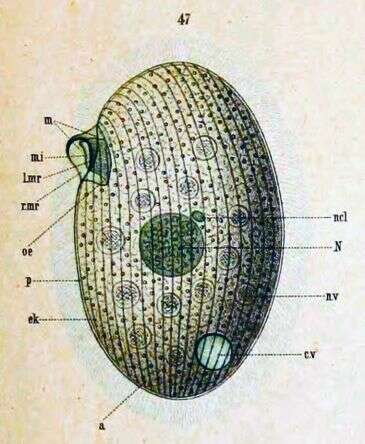
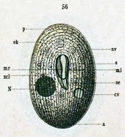
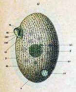
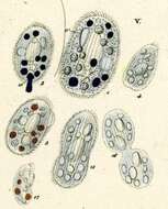
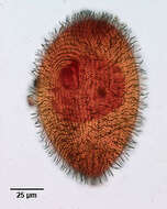
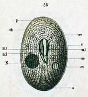
Side view. a -- Anus cv -- Contractile vacuole m -- Undulating membrane mi -- Inner undulating membrane l.mr -- Left membrane edge N -- Macronucleus ncl -- Micronucleus nv -- Food particle oe -- Throat p -- Pellicle r.mr -- Right membrane edge
-
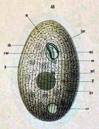
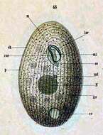
c.v -- Contractile vacuole ek --Ectoplasm l.or -- Left edge of mouth m -- Mouth mi -- inner undulating membrane N -- Macronucleus ncl -- Micronucleus n.v -- Food vacuole oe -- Throat r.or -- Right edge of mouth
-
-
Infraciliature (ventral aspect) of dividing Glaucoma frontata (Stokes,1886) Kahl,1931, a hymenostome ciliate. While most Glaucoma species are ovoid, this elongate species is bluntly rounded anteriorly and tapers posteriorly to rounded tail. Dorsoventrally flattened. The oral aperture is in the anterior third and slightly oblique to the long axis of the body. There is an inconspicuous undulating membrane on the right of the oral aperture and three well developed membranelles. A small rectangular structure at the anterior end of membranelle 2 is termed the âx-bodyâ. The somatic ciliation (about 40 longitudinal kineties) is uniform with a short preoral suture and 5 postoral kineties. The right ventral kineties arch over the cytostome to abut the preoral suture. The forming oral apparatus of the posterior daughter cell (opisthe) partially overlies the macronucleus in this image. Collected from freshwater pond near Boise, Idaho August 2003. Stained by the silver carbonate technique (see Foissner, W.Europ. J. Protistol.27,313-330;1991). Brightfield.
-
Infraciliature of the oral apparatus of of Glaucoma frontata (Stokes,1886) Kahl,1931, a hymenostome ciliate. While most Glaucoma species are ovoid, this elongate species is bluntly rounded anteriorly and tapers posteriorly to rounded tail. Dorsoventrally flattened. The oral aperture is in the anterior third and slightly oblique to the long axis of the body. There is an inconspicuous undulating membrane on the right of the oral aperture and three well developed membranelles. A small rectangular structure at the anterior end of membranelle 2 is termed the "x-body" (seen here). The somatic ciliation (about 40 longitudinal kineties) is uniform with a short preoral suture and 5 postoral kineties. The right ventral kineties arch over the cytostome to abut the preoral suture. The ovoid macronucleus and single micronucleus are central. The single contractile vacuole is located laterally in the midbody. Collected from freshwater pond near Boise, Idaho August 2003. Stained by the silver carbonate technique (see Foissner, W.Europ. J. Protistol.27,313-330;1991). Brightfield.
-
Infraciliature (dorsal aspect) of Glaucoma frontata (Stokes,1886) Kahl,1931, a hymenostome ciliate. While most Glaucoma species are ovoid, this elongate species is bluntly rounded anteriorly and tapers posteriorly to rounded tail. Dorsoventrally flattened. The oral aperture is in the anterior third and slightly oblique to the long axis of the body. There is an inconspicuous undulating membrane on the right of the oral aperture and three well developed membranelles. A small rectangular structure at the anterior end of membranelle 2 is termed the âx-bodyâ. The somatic ciliation (about 40 longitudinal kineties) is uniform with a short preoral suture and 5 postoral kineties. The right ventral kineties arch over the cytostome to abut the preoral suture. The ovoid macronucleus and single micronucleus are central. The single contractile vacuole is located laterally in the midbody. Collected from freshwater pond near Boise, Idaho August 2003. Stained by the silver carbonate technique (see Foissner, W.Europ. J. Protistol.27,313-330;1991). Brightfield.
-

Infraciliature (ventral aspect) of Glaucoma frontata (Stokes,1886) Kahl,1931, a hymenostome ciliate. While most Glaucoma species are ovoid, this elongate species is bluntly rounded anteriorly and tapers posteriorly to rounded tail. Dorsoventrally flattened. The oral aperture is in the anterior third and slightly oblique to the long axis of the body. There is an inconspicuous undulating membrane on the right of the oral aperture and three well developed membranelles. A small rectangular structure at the anterior end of membranelle 2 is termed the âx-bodyâ. The somatic ciliation (about 40 longitudinal kineties) is uniform with a short preoral suture and 5 postoral kineties. The right ventral kineties arch over the cytostome to abut the preoral suture. The ovoid macronucleus and single micronucleus are central. The single contractile vacuole is located laterally in the midbody. Collected from freshwater pond near Boise, Idaho August 2003. Stained by the silver carbonate technique (see Foissner, W.Europ. J. Protistol.27,313-330;1991). Brightfield.
-
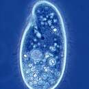
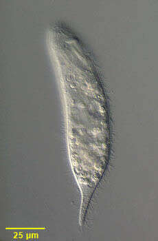
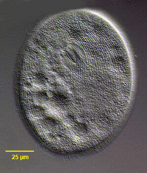
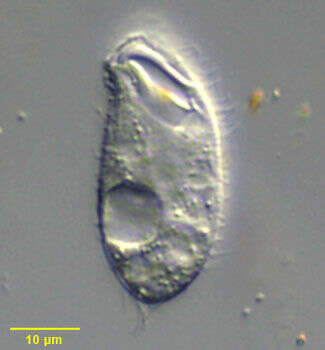
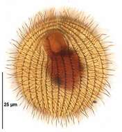
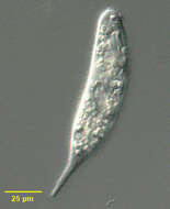
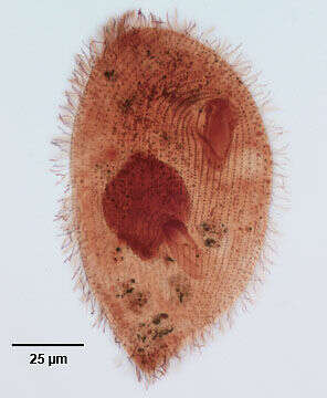
Portrait of Glaucoma frontata (Syn. Dallasia frontata), a hymenostome ciliate. While most Glaucoma species are ovoid, this elongate species is bluntly rounded anteriorly and tapers posteriorly to rounded tail. Dorsoventrally flattened. The oral aperture is in the anterior third and oblique to the long axis of the body. There is an inconspicuous undulating membrane on the right of the oral aperture and three well developed membranelles. The somatic ciliation is uniform with a short preoral suture and 5 or more postoral kineties. The ovoid macronucleus and single micronucleus are central. The single contractile vacuole is located laterally in the midbody. Numerous food vacuoles are visible in this image. Mainly bactivorous. Collected from freshwater pond near Boise, Idaho August 2003. DIC optics.
-
Portrait of Glaucoma frontata (Syn. Dallasia frontata), a hymenostome ciliate. While most Glaucoma species are ovoid, this elongate species is bluntly rounded anteriorly and tapers posteriorly to rounded tail. Dorsoventrally flattened. The oral aperture is in the anterior third and oblique to the long axis of the body. There is an inconspicuous undulating membrane on the right of the oral aperture and three well developed membranelles. The somatic ciliation is uniform with a short preoral suture (seen in this image) and 5 or more postoral kineties. The ovoid macronucleus and single micronucleus are central. The single contractile vacuole is located laterally in the midbody. Numerous food vacuoles are visible in this image. Mainly bactivorous. Collected from freshwater pond near Boise, Idaho August 2003. DIC optics
-
a -- Anus cv -- Contractile vacuole ek -- Ectoplasm ml -- Left undulating membrane mr -- Right undulating membrane N -- Macronucleus ncl -- Micronucleus nv -- Food vacuole o -- Mouth oe -- Throat p -- Pellicle
-
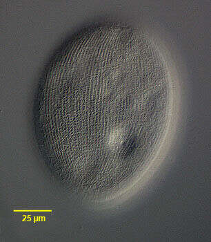
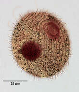
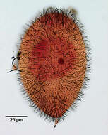
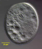
Ventral view of Epenardia myriophylli (Penard, 1922) Corliss, 1971, a large glaucomid ciliate. This cell is slighlty compressed. The body is ellipsoid with minimal dorsoventral flattening. The relatively small obliquely oriented cytostome is in the anterior 1/4. The three adoral membranelles are located in the deep buccal cavity. There is an undulating membrane on the right. E. myriophylli differs from Glaucoma species by its dense (80-110) somatic kineties and the broad M3 which is wider than M2 and a preoral suture in the long axis. There are approximately 9 postoral kineties. The spherical macronucleus and micronucleus are centrally located. The contractile vacuole empties through a single excretory pore on the right dorsal surface. The pellicle is pitted with regular square depressions. Collected from a freshwater drainage ditch near Boise, Idaho. April 2005. DIC
-
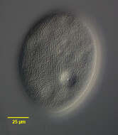
Dorsal view of Epenardia myriophylli (Penard, 1922) Corliss, 1971, a large glaucomid ciliate. This cell is slighlty compressed. The body is ellipsoid with minimal dorsoventral flattening. The relatively small obliquely oriented cytostome is in the anterior 1/4. The three adoral membranelles are located in the deep buccal cavity. There is an undulating membrane on the right. E. myriophylli differs from Glaucoma species by its dense (80-110) somatic kineties and the broad M3 which is wider than M2 and a preoral suture in the long axis. There are approximately 9 postoral kineties. The spherical macronucleus and micronucleus are centrally located. The contractile vacuole empties through a single excretory pore on the right dorsal surface. The pellicle is pitted with regular square depressions. Collected from a freshwater drainage ditch near Boise, Idaho. April 2005. DIC
-
Oral infraciliature (ventral view) of Epenardia myriophylli (Penard, 1922) Corliss, 1971, a large glaucomid ciliate. This cell is slighlty compressed. The body is ellipsoid with minimal dorsoventral flattening. The relatively small obliquely oriented cytostome is in the anterior 1/4. The three adoral membranelles are located in the deep buccal cavity. There is an undulating membrane on the right. E. myriophylli differs from Glaucoma species by its dense (80-110) somatic kineties and the broad M3 which is wider than M2 and a preoral suture in the long axis. There are approximately 9 postoral kineties. The spherical macronucleus and micronucleus are centrally located. The contractile vacuole empties through a single excretory pore on the right dorsal surface. The pellicle is pitted with regular square depressions. Collected from a freshwater drainage ditch near Boise, Idaho. April 2005.Silver carbonate stain (see Foissner, W. Europ. J. Protistol., 27:313-330;1991). Brightfield.
-
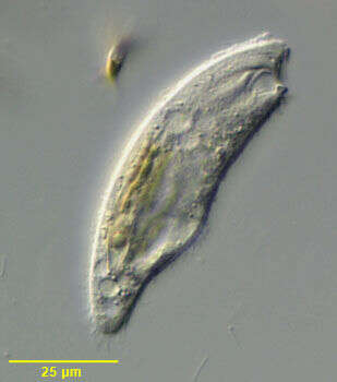
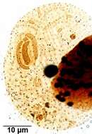
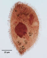
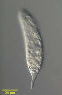
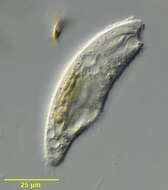
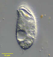
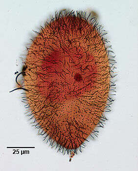
Portrait of the glaucomid ciliate, Espejoia mucicola. Espejoia is found in two morphologic forms, the macrostome (pictured here) and the microstome. Macrostome individuals are larger and the oral aperture occupies the anterior quarter of the cell often reaching the anterior apex. There are three well-developed oral membranelles. The macronucleus is round to irregular. The contractile vacuole is located in the posterior 1/3. Somatic ciliation is uniform with a preoral suture in the microstome form. E. mucicola is often found in association with the mucus coating of dipteran egg cases and the mucilaginous coat of algal thalli. Collected from slow moving freshwater stream near Boise, Idaho in May 2003. DIC optics.
-
Portrait of the glaucomid ciliate, Espejoia mucicola. Espejoia is found in two morphologic forms, the macrostome (pictured here) and the microstome. Macrostome individuals are larger and the oral aperture occupies the anterior ? of the cell often reaching the anterior apex. There are three well-developed oral membranelles. The macronucleus is round to irregular. The contractile vacuole is located in the posterior 1/3. Somatic ciliation is uniform with a preoral suture in the microstome form. E. mucicola is often found in association with the mucus coating of dipteran egg cases and the mucilaginous coat of algal thalli. Collected from slow moving freshwater stream near Boise, Idaho in May 2003. DIC optics.
-
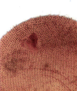
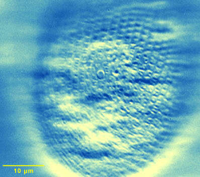
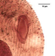
Epenardia (named after Eugene Penard) is a oligohymenophoran ciliate with a subapical mouth, body densely ciliated. This detail shows the cortex in the region of the contractile vacuole, showing the pore through which the fluid is periodically discharged. The cortex is pitted at the site where the cilia emerge.