-
All Biocode files are based on field identifications to the best of the researcher’s ability at the time.
-
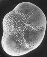
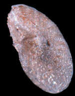
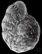
Foraminiferans living in polluted environments often show alterations in the morphology of their tests. This individual, isolated from a site in Norway which is contaminated with heavy metals, exhibits reduced chamber size in some of its chambers (notice that the test is not evenly rounded.) Image courtesy of Dr. Elisabeth Alve, University of Oslo. Citation: Alve, E. Benthic foraminifera reflecting pollution. Journal of Foraminiferal Research 21:1-19.
-
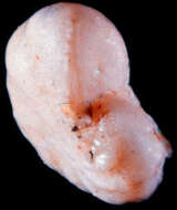
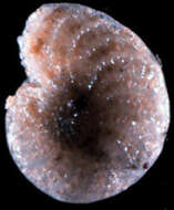
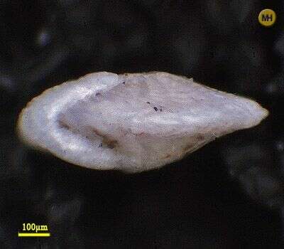
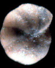
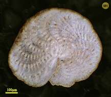
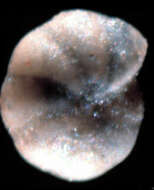
Image of the holotype. Specimen is badly etched and difficult to place taxonomically. Test is 1.18 mm. in long dimension. Image courtesy of David B. Scott, Dalhousie University. This image was originally published in
Palaeologica Electronica, vol. 3, issue 2, and is used with the kind permission of that journal and the Paleontological Association.
-
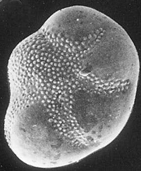
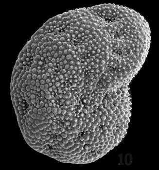
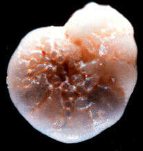
Cribroelphidium poeyanum, an epiphytic foraminiferan from Twin Cays, Belize
-
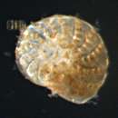
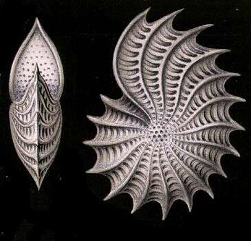
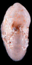
Polystomella aculeata.
-
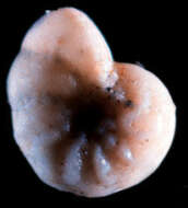
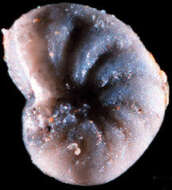
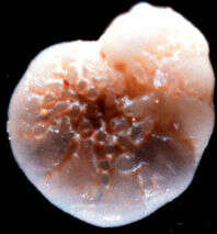
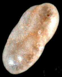
Image of the holotype. Test is 1.26 mm. across. Image courtesy of David B. Scott, Dalhousie University. This image was originally published in
Palaeologica Electronica, vol. 3, issue 2, and is used with the kind permission of that journal and the Paleontological Association.
-
This unidentified species inhabited the Laguna Madre area at one time, but is not found there now. Image courtesy of Pamela Stephens, Midwestern State University.
-
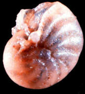
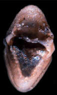
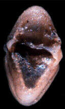
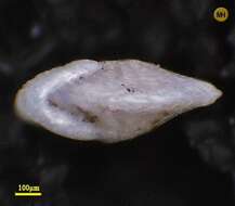
Image of holotype, showing the aperture. Image courtesy of David B. Scott, Dalhousie University. This image was originally published in
Palaeologica Electronica, vol. 3, issue 2, and is used with the kind permission of that journal and the Paleontological Association.
-
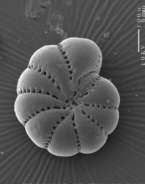
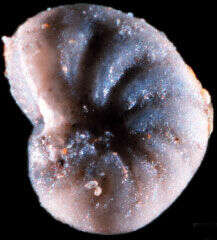
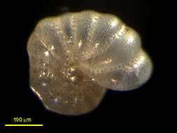
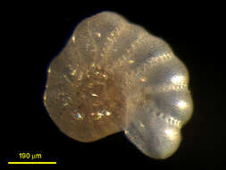
Dorsal view of Elphidion. Collected from Eel Pond, Woods Hole. Image by L Wegener, DJ Patterson and D Lahr.
-
Image of holotype. Test is 1.1 mm. across. Image courtesy of David B. Scott, Dalhousie University. This image was originally published in
Palaeologica Electronica, vol. 3, issue 2, and is used with the kind permission of that journal and the Paleontological Association.
-
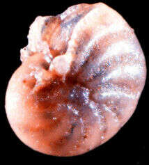
Ventral view. This foram was collected by L. Wegener from Eel Pond, Woods Hole. Image by L Wegener, DJ Patterson and D Lahr.
-
Image of the holotype. The test is etched, and the orange encrustations are not part of the original test. Image courtesy of David B. Scott, Dalhousie University. This image was originally published in
Palaeologica Electronica, vol. 3, issue 2, and is used with the kind permission of that journal and the Paleontological Association.
-
From Laguna Madre, Texas. Image courtesy of Pamela Stephens, Midwestern State University.
-
Image of the holotype. The test is 1.2 mm. across. Image courtesy of David B. Scott, Dalhousie University. This image was originally published in
Palaeologica Electronica, vol. 3, issue 2, and is used with the kind permission of that journal and the Paleontological Association.
-
Sample collected at Hamble Estuary, Hampshire, England. Image courtesy of Elisabeth Alve, University of Oslo. Originally published in the Journal of Foraminiferal Research 31:1; used with permission.
-
Image of the holotype. The youngest chamber is broken on this specimen, which is 1.33 mm. across. Image courtesy of David B. Scott, Dalhousie University. This image was originally published in
Palaeologica Electronica, vol. 3, issue 2, and is used with the kind permission of that journal and the Paleontological Association.
-
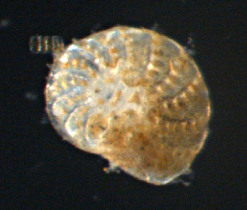
Smoegen/Skagerrak/Sweden, 58.35 N, 11.22 E found at beach
-
Image of the holotype. The remains of the aperture of this broken test face toward the viewer. Image courtesy of David B. Scott, Dalhousie University. This image was originally published in
Palaeologica Electronica, vol. 3, issue 2, and is used with the kind permission of that journal and the Paleontological Association.
-
Smoegen/Skagerrak/Sweden, 58.35 N, 11.22 E found at beach.
-
Image of holotype. Specimen is broken an etched. Test is 1.41 mm. across. Image courtesy of David B. Scott, Dalhousie University. This image was originally published in
Palaeologica Electronica, vol. 3, issue 2, and is used with the kind permission of that journal and the Paleontological Association.
-
Image of the holotype, with the aperture toward the viewer. Image courtesy of David B. Scott, Dalhousie University. This image was originally published in
Palaeologica Electronica, vol. 3, issue 2, and is used with the kind permission of that journal and the Paleontological Association.
-
Discription to come
-
Image of the holotype. The test, which is in poor condition, is 0.47 mm. across. Image courtesy of David B. Scott, Dalhousie University. This image was originally published in
Palaeologica Electronica, vol. 3, issue 2, and is used with the kind permission of that journal and the Paleontological Association.
-
Image courtesy of David B. Scott, Dalhousie University. This image was originally published in
Palaeologica Electronica, vol. 3, issue 2, and is used with the kind permission of that journal and the Paleontological Association.