-
-
Notostomus robustus, sp. nov. Lateral view of female, from Station 2074.
-
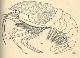
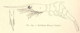
Systellaspis Bouvieri Coutiere.
-
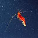
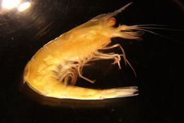
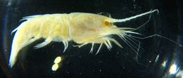
Systellaspis braueri from 900 m depth 100 mi W of Point Conception, CA (Photo by: Dave Cowles April 1996)
-
Whole individual. Carapace length 1.9 cm. Notice how the 6th abdominal segment is twice as long or more than the 5th abdominal segment, and that it does not have a dorsal carina. Also note that abdominal segments 3, 4, and 5 have a posteromesial spine.
-
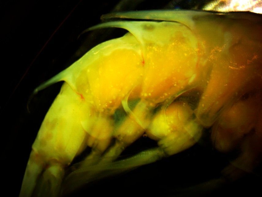
This photograph of the left side of the shrimp shows pereopods 3-5 (#5 is to the right). The endopods are robust and have reddish setae. The ventral edge of the carapace is visible at the top of the photo. The epipod on pereopod 4 can be seen must below the edge of the carapace, branching from the coxa. The exopods, which look like long flexible yellowish extensions, can be seen on pereopods 3-5, branching from the basis. Note that the exopods are of similar length to each other.
-
Lateral view of abdominal segments 2-3. Segment 2 has no dorsal carina nor posteromesial spine, while segment 3 has both a carina and a spine.
-
A side view of abdominal segments 1-3. Note that segment 2 has no posterior dorsal spine, while segment 3 is dorsally carinate and ends with a long medial dorsal spine. This view is of the left side of the shrimp, anterior is to the left.
-
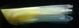
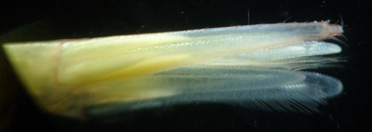
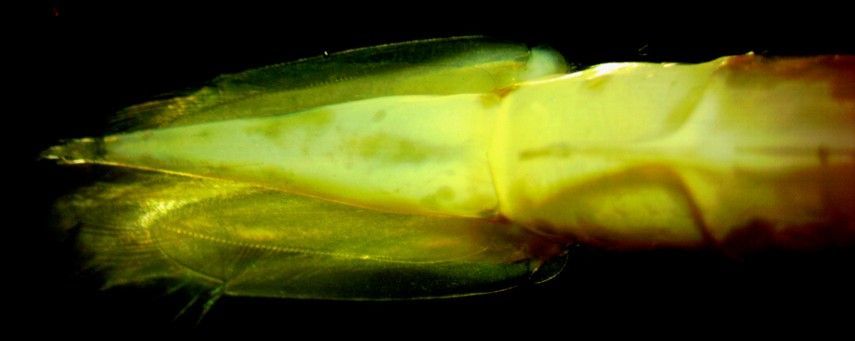
Dorsal (slight angle) view of the uropods and telson. The spiny endpiece on the telson has been slightly damaged but the long lateral spines at the base can still be seen. A few of the dorsolateral spines along the telson can also be seen along the top margin.
-
A side view of abdominal segments 4-6 (4 is to the left). Note the small posterior medial dorsal spines on segments 4,5 and the small spines lateral to them. Note also the tooth on the posterior margin of the pleuron of segment 5. Anterior is to the left.
-
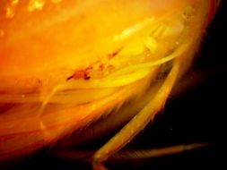
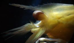
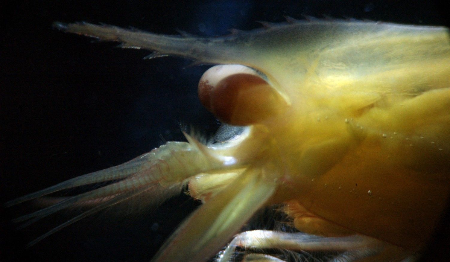
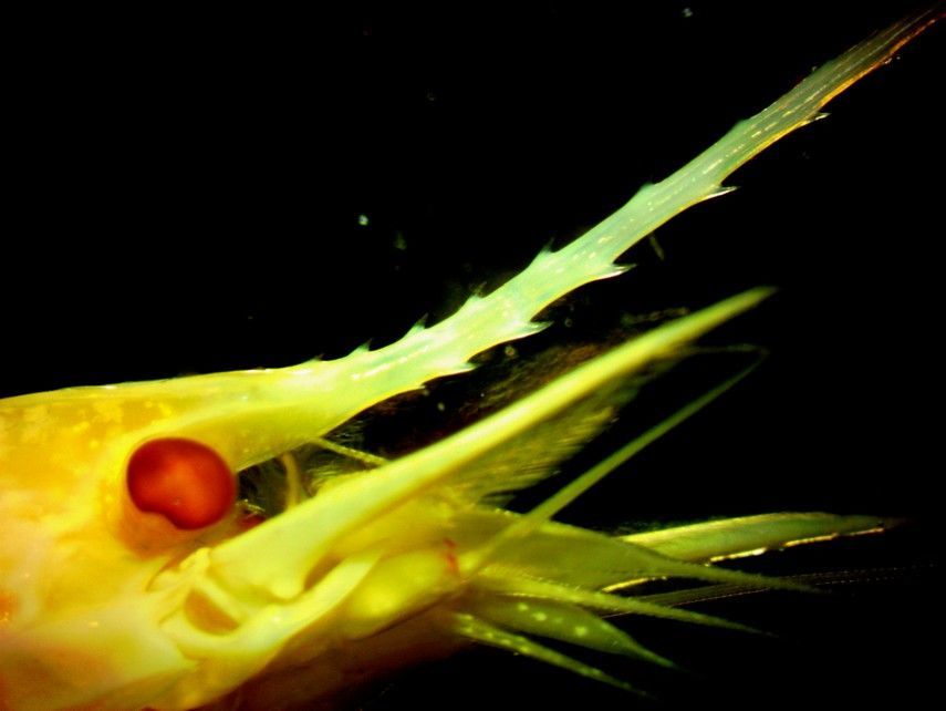
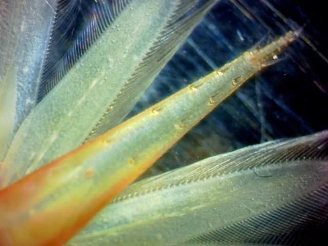
This side view of the head shows the long, toothed rostrum which extends out to near the end of the antennal scales (which are deflected downward out of view in this photo)
-
This dorsal view of abdominal segments 5-6 shows the small posterior medial dorsal spine on segment 5 and the small spines lateral to it. Anterior is to the left.
-
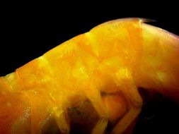
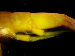
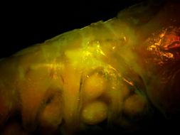
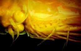
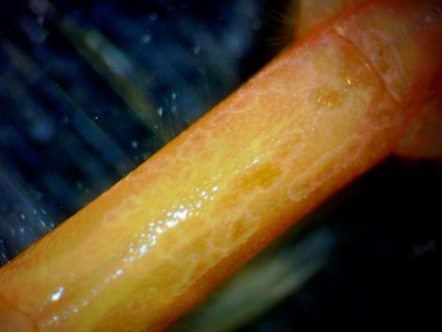
The three characteristic carinae which run nearly to the posterior end of the carapace can be seen in this side view. Dorsally there is a carina which runs from the rostrum (left) to near the posterior end of the carapace on the right. A small, sinuous carina runs from near the eye along the mid-side of the carapace. In this photo a number of bubbles are adhered to it. A sharp carina lines the ventral margin of the carapace. In this preserved specimen the ventral carina has retained much of its red color while the rest of the carapace has faded to yellow.
-
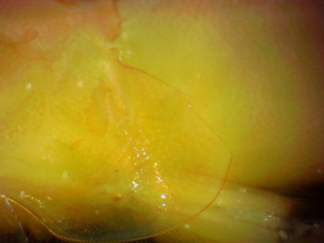
This lateral closeup view of abdominal pleuron 5 shows the sharp tooth along the posterior margin. Anterior is to the left.
-
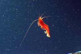
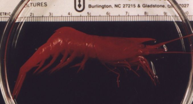
Systellaspis cristata, caught off Point Conception, CA at 600-750 m depth. (Photo by: Dave Cowles, Sept 1995)
-
This dorsal view of abdominal segment 6 shows that it rounded and has no dorsal carina (ridge). Anterior is to the left and the base of the uropods and telson are on the upper right.
-
As with other Oplophorus species, O. spinosus has a long rostrum with many spines on both the dorsal and ventral surface. Note the well-developed eye as well.
-
This dorsal view of the pleopods and telson show that the telson has a shallow dorsal groove (sulcus). It also has a spiny endpiece flanked by two spines, and two dorsolateral rows of 4-8 spines.
-
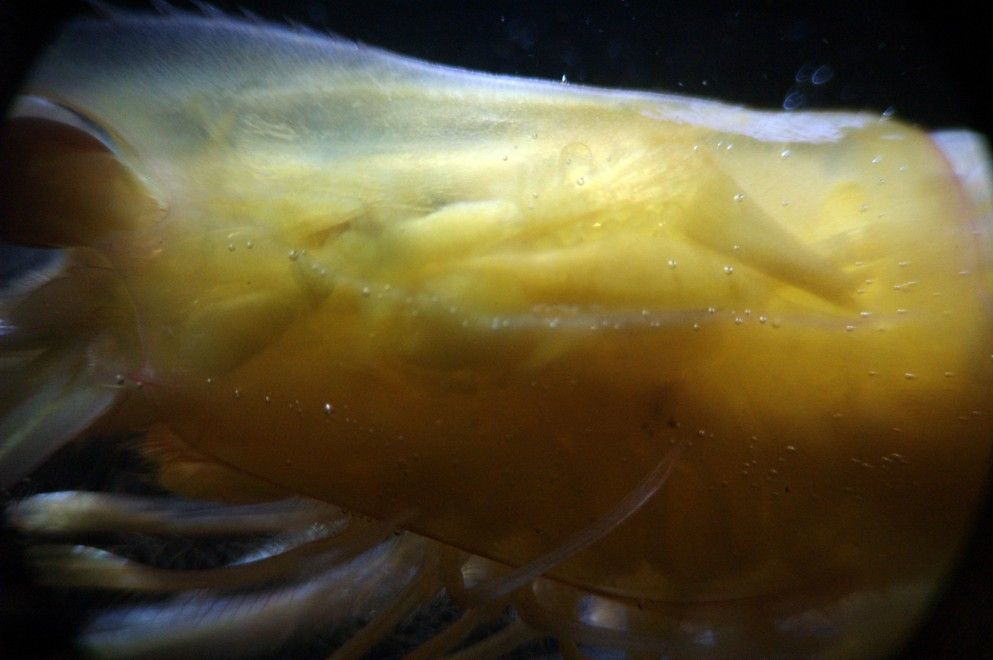
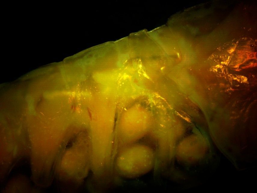
This right-side view of the anterior (first 3) abdominal segments (the posterior thorax is at the right side) shows that the second abdominal segment does not have a large posterior mid-dorsal spine. The large eggs can be seen attached to the pleopods.
-
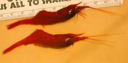
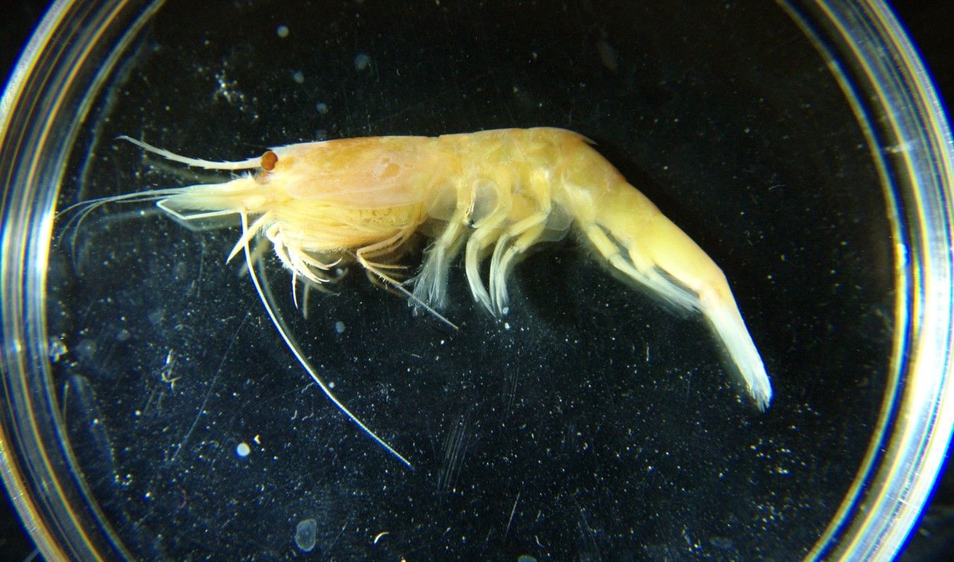
Systellaspis debilis, preserved specimen captured in midwater off Oahu, Hawaii in 1996. Compare petri dish for size. In life the shrimp would be bright red. (Photo by: Dave Cowles)
-
This right-side view of the posterior abdominal segments show the large posterior-pointing mid-dorsal spines on several of them. Abdominal segment 3 is at the top right, then segments 4,5,6, and the base of the telson is visible at the bottom left. Note that segment 6 is not dorsally carinate.
-
Dorsal view of the rear abdomen and telson. The rightmost segment is abdominal segment 5. Segment 6 is below the posterior spine from segment 5. To the left are the uropods and telson.
-
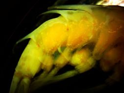
In O. spinosus there is no sharp tooth on the posterior margin of the ventralcarapace. In this view of the animal's right side, the ventral margin of the carapace plus the pereopods can be seen to the right and the first abdominal segment with pleopods and several eggs is visible to the left. The posterior margin of the ventralcarapace has an acutely rounded corner but no sharp tooth.
-
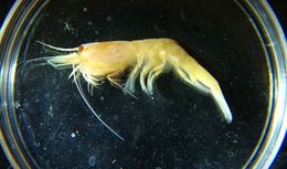
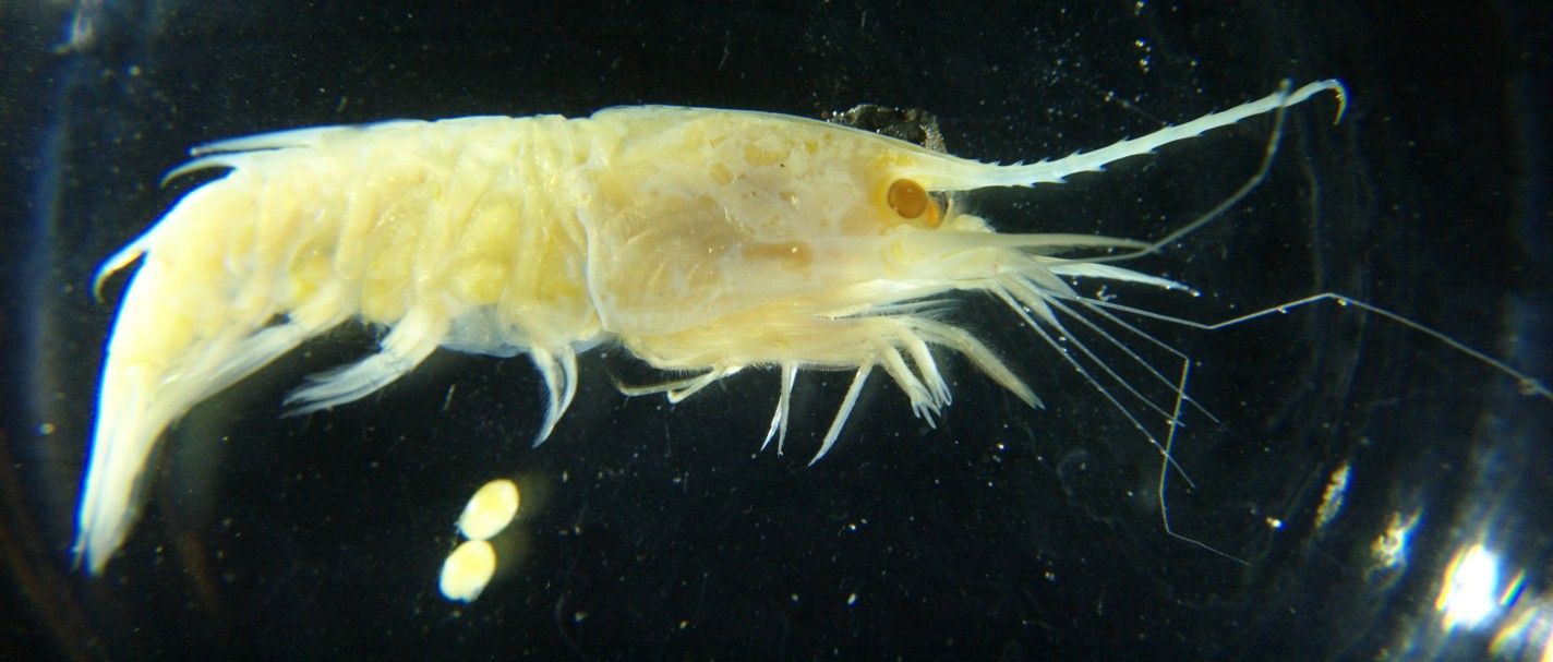
A female Oplophorus spinosus carrying eggs, carapace length 1.5 cm, captured in midwater off Hawaii in 1996. In real life this species would be partly pinkish-red and partly transparent. The specimen above has been preserved in formalin. Several eggs have dropped off the pleopods. (Photo by: Dave Cowles, 2012 )