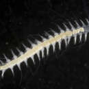
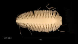
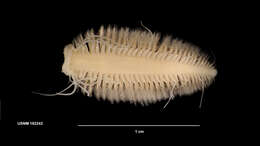
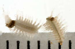
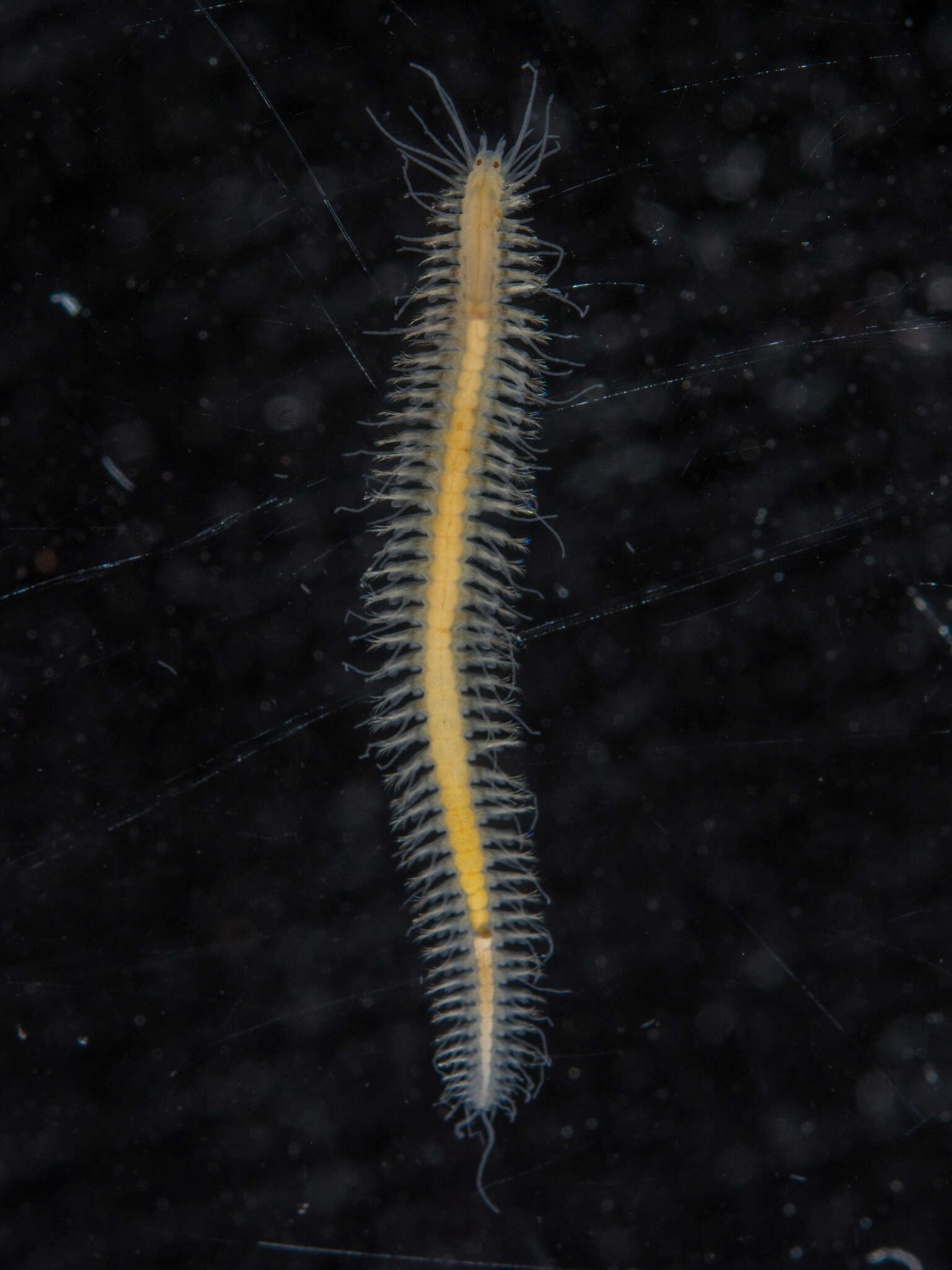
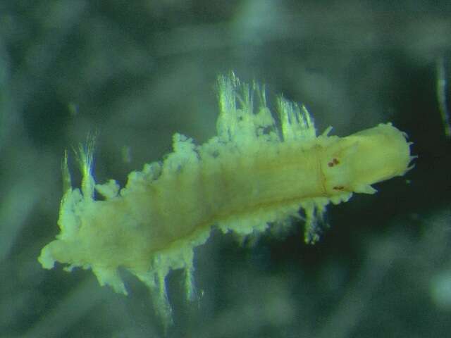
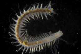
Collected from Puget Sound sediments and photographed by the Washington State Department of Ecologys Marine Sediment Monitoring Team. For more information about this teams work visit: www.ecy.wa.gov/programs/eap/psamp/index.htm.
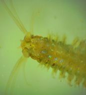
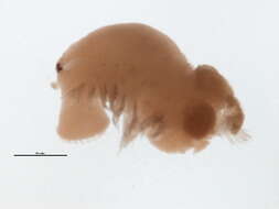
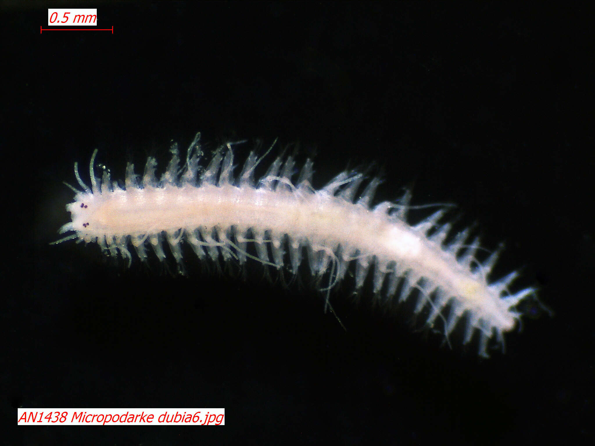
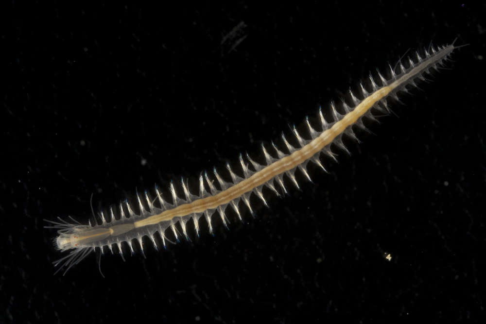
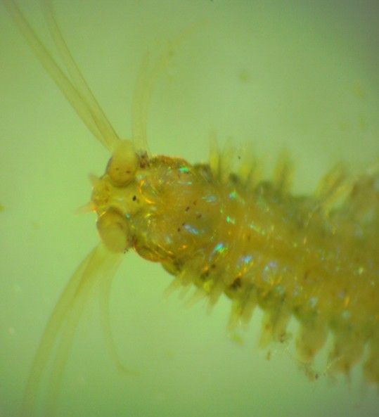
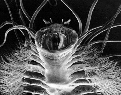
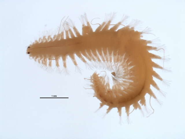
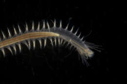
Dorsal view of the head. Note the 4 dorsal eyes and the two 2-segmented prostomial palps with the two segments of similar length. The two prostomial antennae can also be partly seen anteriorly.
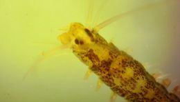
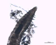
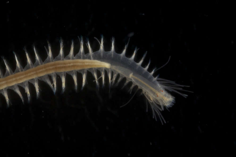
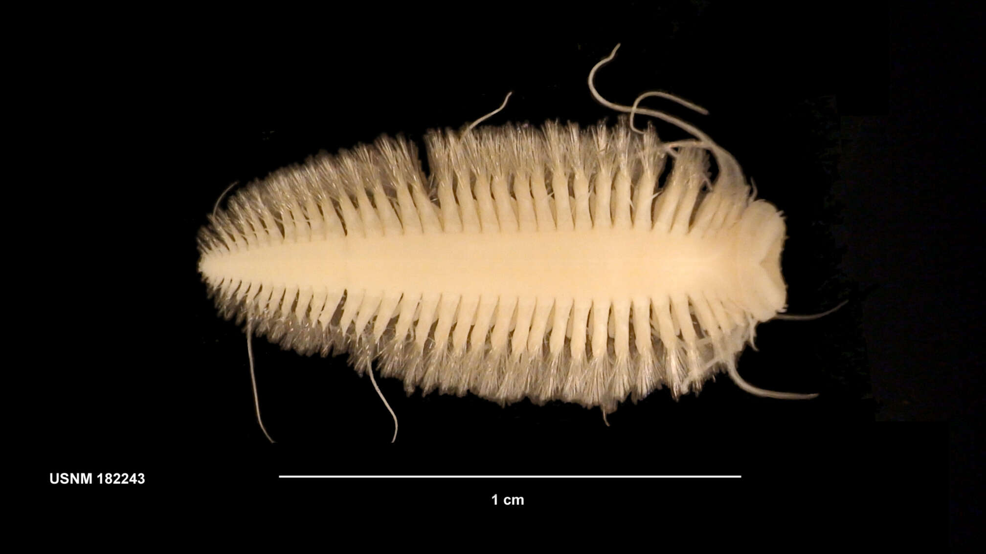
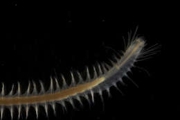
This ventral view of the head shows the open mouth in the peristomium. The pharynx is inside the mouth. This species has no teeth on its pharynx, although this would not be visible unless the animal everted its pharynx. The two prostomial antennae can also be seen. The larger, 2-segmented palps are extending from the prostomium directly toward the viewer. This view of the head (top right) and pygidium (left) shows that the pygidium has two long projecting cirri.
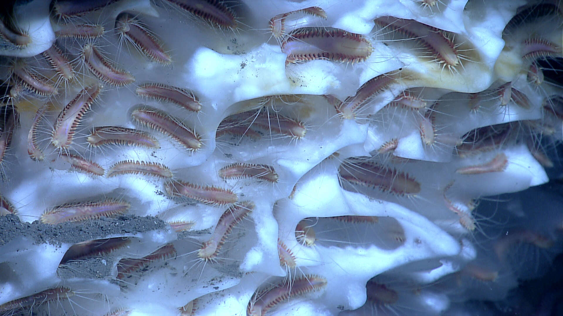
Description: English: Ice Worm ; Source ; Ice Worm Photo courtesy of NASA JPL Français : Vers vivant sur les hydrates de méthane, à grande profondeur (trouvé pour la 1ère fois en 1987 dans le Golfe du Mexique). Ce vers semble capable de coloniser la surface du méthane hydraté, où il vivrait en y mangeant les bactéries méthanotrophe qu'il y trouve. Il est pour cette raison nommé vers de glace (Ice worm) par les anglophones. Date: import on commons : 2010/11/03. Source: Source ; Ice Worm Photo courtesy of NASA JPL. Author: NASA.
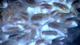
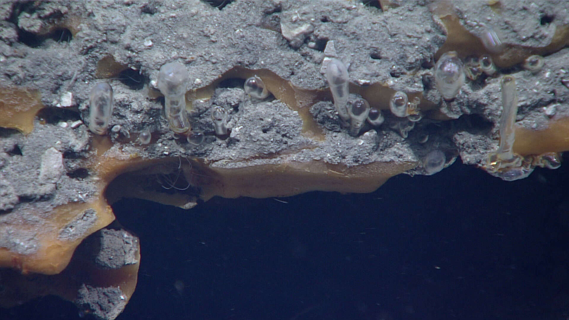
Description: English: Pink “ice worms” are visible beneath the overhang in the center left part of this photo. Ice worms were also seen in some of the burrows in the surrounding gas hydrate, which appears orange due to impurities. Active methane emissions occur from beneath the ledge, through conduits at the base of the inverted droplets attached to the sediment and through a bubble tube at the right of the image. Both the bubble tubes and the inverted droplets are encased in clear gas hydrate. Source: https://oceanexplorer.noaa.gov/okeanos/explorations/ex1711/logs/dec20/media/iceworms.html. Author: Image courtesy of the NOAA Office of Ocean Exploration and Research, Gulf of Mexico 2017.