-
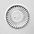
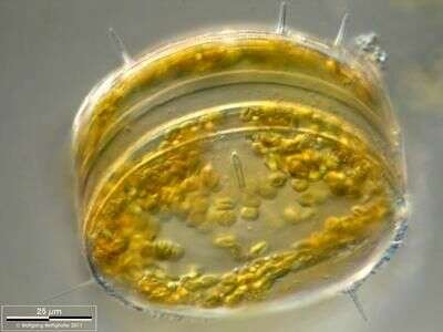
Thalassiosira punctigera.Some specimen of this centric diatom carried naviculoid ones on the valve(s). Scale bar indicates 25 m. The image was built up using several photomicrographic frames with manual stacking technique. Sample from North Sea near Heligoland (spring diatom bloom). Images were taken using Zeiss Universal with Olympus C7070 CCD camera.For more look at
www.protisten.de/english/gallery_main/gallery_main.htmlFor high-resolution images please ask postmaster@protisten.de.
-
Grove, O, Galicia, Spain
-
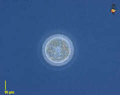
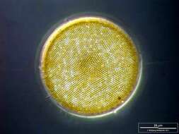
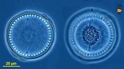
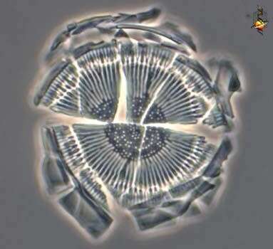
Centric diatom, seen from valve view. The pattern of pores in the frustule is used in identification. Marine. Phase contrast.
-
-
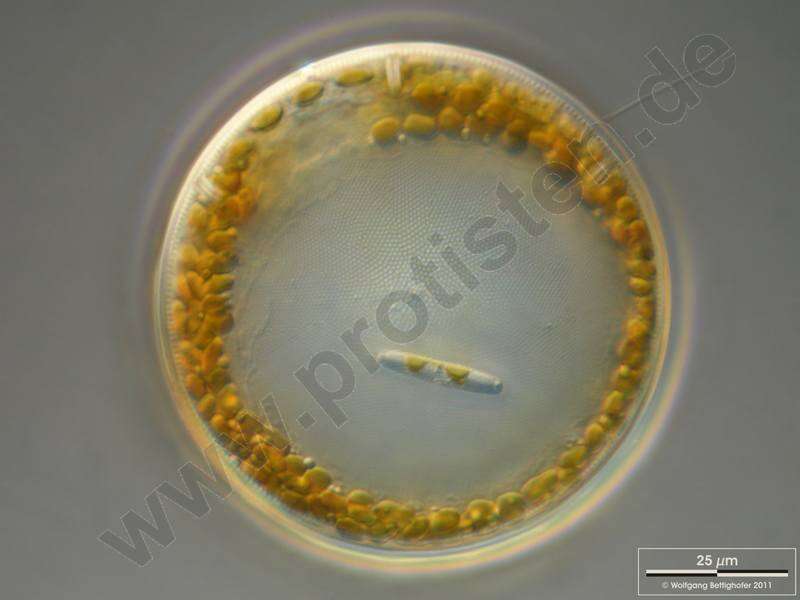
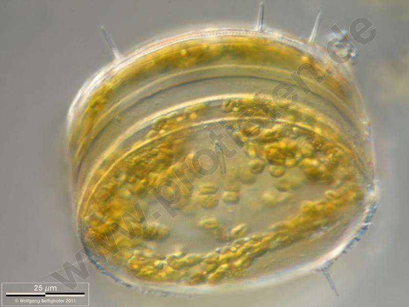
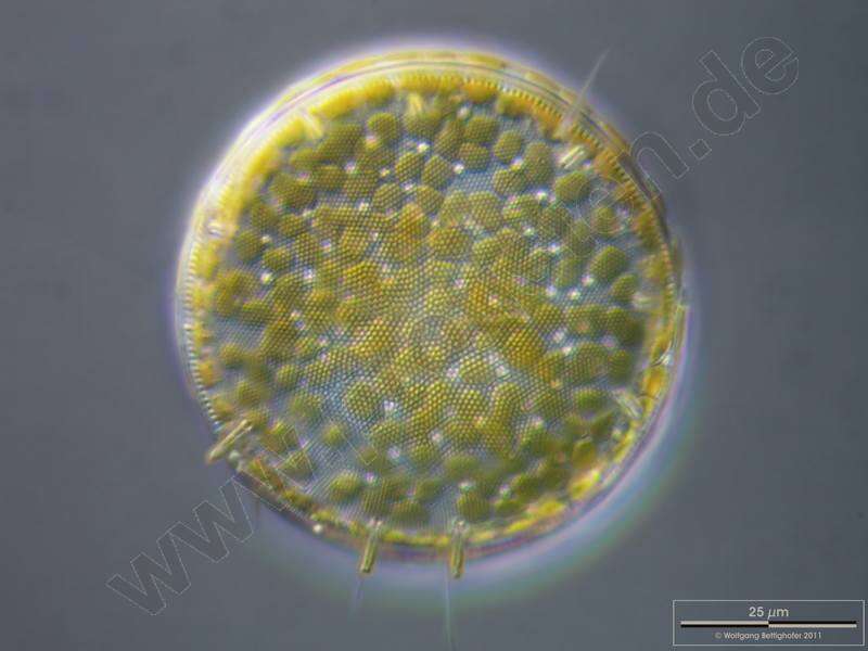
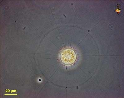
The oblique view exhibits short silicous spines, the so called occluded processes. On the lower left near the scale bar a chitinous spine is visible. Scale bar indicates 25 µm. The image was built up using several photomicrographic frames with manual stacking technique. Sample from North Sea near Heligoland (spring diatom bloom). Images were taken using Zeiss Universal with Olympus C7070 CCD camera.
-
Thalassiosira punctigera.The oblique view exhibits short silicous spines, the so called occluded processes. On the lower left near the scale bar a chitinous spine is visible. Scale bar indicates 25 m. The image was built up using several photomicrographic frames with manual stacking technique. Sample from North Sea near Heligoland (spring diatom bloom). Images were taken using Zeiss Universal with Olympus C7070 CCD camera.For more look at
www.protisten.de/english/gallery_main/gallery_main.htmlFor high-resolution images please ask postmaster@protisten.de.
-
Grove, O, Galicia, Spain
-
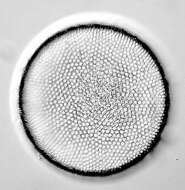
Cyclotella (sike-low-tell-a). Centric diatom, frustule only, seen from valve view, with the two frustules seen at slightly different focal planes. . The pattern of pores in the frustule is used in identification. Phase contrast.
-
One labiate process is visible together with some chitinous spines. Scale bar indicates 25 µm. The image was built up using several photomicrographic frames with manual stacking technique. Sample from North Sea near Heligoland (spring diatom bloom). Images were taken using Zeiss Universal with Olympus C7070 CCD camera.
-
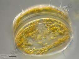
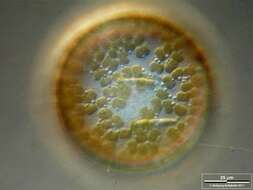
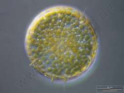
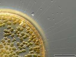
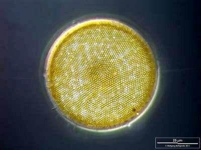
Some specimen of this centric diatom carried naviculoid ones on the valve(s). Scale bar indicates 25 µm. The image was built up using several photomicrographic frames with manual stacking technique. Sample from North Sea near Heligoland (spring diatom bloom). Images were taken using Zeiss Universal with Olympus C7070 CCD camera.
-
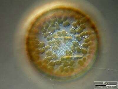
Thalassiosira punctigera.Silicious processes (the labiate and the occluded ones) are visible. Scale bar indicates 25 m. The image was built up using several photomicrographic frames with manual stacking technique. Sample from North Sea near Heligoland (spring diatom bloom). Images were taken using Zeiss Universal with Olympus C7070 CCD camera.For more look at
www.protisten.de/english/gallery_main/gallery_main.htmlFor high-resolution images please ask postmaster@protisten.de.
-
Grove, O, Galicia, Spain
-
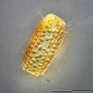
Cyclotella (sike-low-tell-a). Centric diatom, seen from valve view. The cell is surrounded by a sheath of mucus. Long thin organic spines project from the cell - and are believed to have a role in flotation. The pattern of pores in the frustule is used in identification. From a freshwater site. Phase contrast.
-
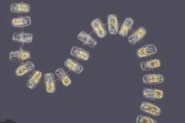
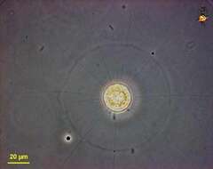
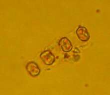
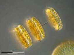
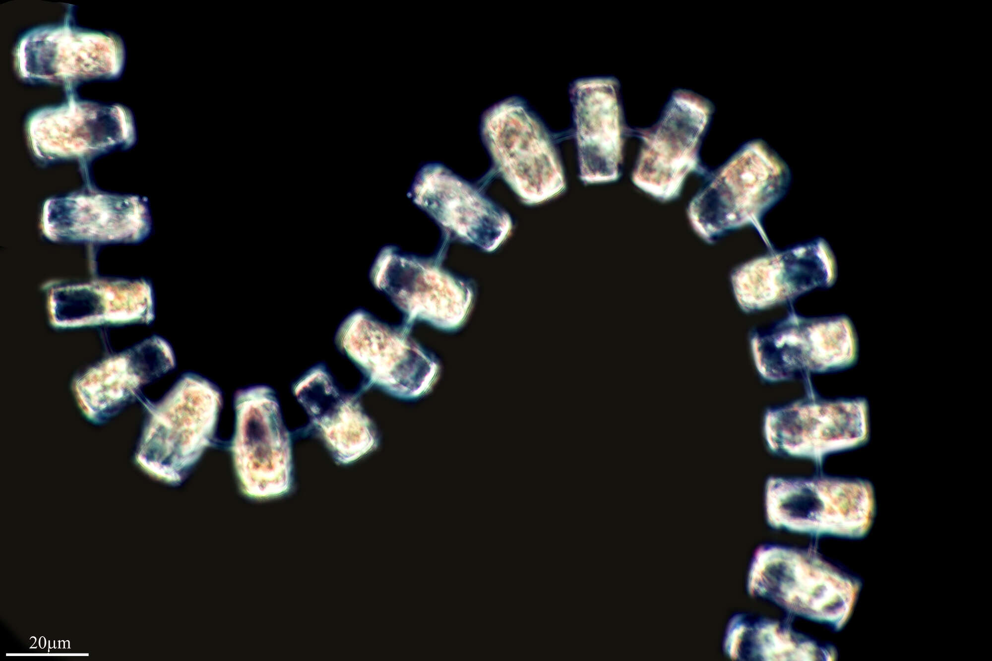
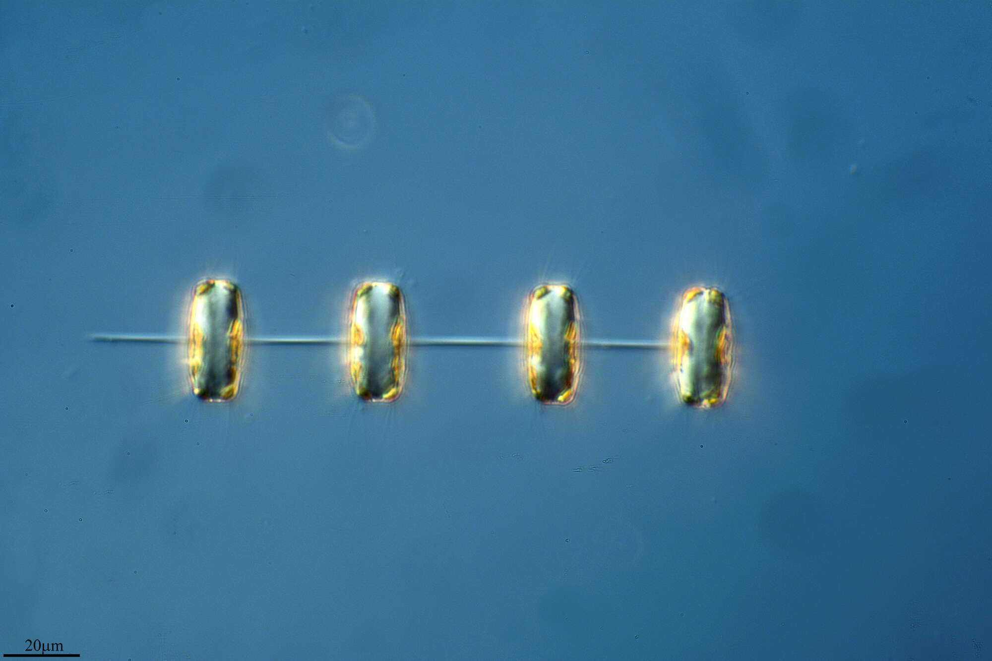
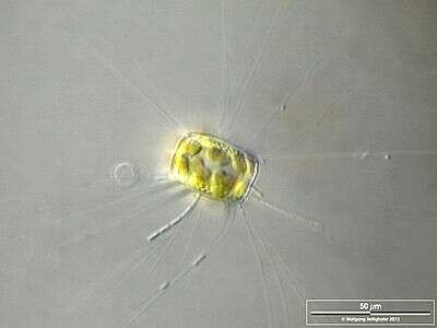
Cells form closely linked straight chains. Long, delicate threads radiate form the valve margins. Valve diameter 34-38 microns.
-
The oblique view exhibits short silicous spines, the so called occluded processes. On the lower left, lower right and central above chitinous spines are visible. Scale bar indicates 50 µm. The image was built up using several photomicrographic frames with manual stacking technique. Sample from North Sea near Heligoland (spring diatom bloom). Images were taken using Zeiss Universal with Olympus C7070 CCD camera.
-
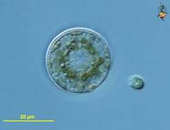
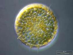
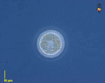
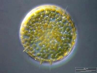
Cyclotella (sike-low-tell-a). Centric diatom, seen from valve view. With many small plastids containing chlorophylls a and c. From a freshwater site. Differential interference contrast.
-
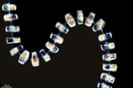
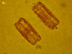
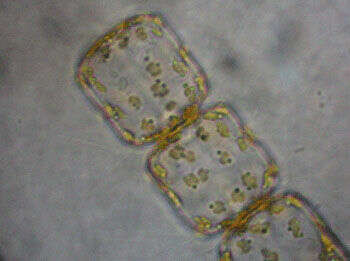
Skeletonema costatum cells are very small and highly variable in shape. They are connected into chains by external tubes of strutted processes. As long as sibling valves or chains are observed this species can be easily identified. However, in chains where the tubes are very short they may be confused with small Thalassiosira species.
-
The oblique view exhibits short silicous spines, the so called occluded processes. Some chitinous spines protruding from the fultoportulae (also called strutted processes) along the dotted valve margin are also visible. Scale bar indicates 50 µm. The image was built up using several photomicrographic frames with manual stacking technique. Sample from North Sea near Heligoland (spring diatom bloom). Images were taken using Zeiss Universal with Olympus C7070 CCD camera.
-
Cyclotella (sike-low-tell-a). Centric diatom, frustule only, seen from valve view, frustule broken from cover-slip pressure to show the brittle nature of the frustule. The pattern of pores in the frustule is used in identification. Phase contrast.
-
-
Silicious processes (the labiate and the occluded ones) are visible. Scale bar indicates 25 µm. The image was built up using several photomicrographic frames with manual stacking technique. Sample from North Sea near Heligoland (spring diatom bloom). Images were taken using Zeiss Universal with Olympus C7070 CCD camera.
-
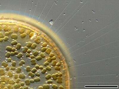
The thin, barely visible floating extensions are made of chitin. Furthermore, filamentous bacteria colonies are attached. Scale bar indicates 50 µm. Sample from the Lake Constance (vicinity of Bodman). The image was built up using several photomicrographic frames with manual stacking technique. Images were taken using Zeiss Universal with Olympus C7070 CCD camera.Image under Creative Commons License V 3.0 (CC BY-NC-SA).
-
The members of the colony are interconnected with a bundle of threads. Numerous delicate spines protruding from the valve's margin are visible. Scale bar indicates 50 µm. The image was built up using several photomicrographic frames with manual stacking technique. Sample from North Sea near Heligoland (spring diatom bloom). Images were taken using Zeiss Universal with Olympus C7070 CCD camera.
-
The image shows numerous chitinous spines which minimize their sedimentation speed. Scale bar indicates 25 µm. The image was built up using several photomicrographic frames with manual stacking technique. Sample from North Sea near Heligoland (spring diatom bloom). Images were taken using Zeiss Universal with Olympus C7070 CCD camera.Image under Creative Commons License V 3.0 (CC BY-NC-SA).