-
-
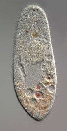
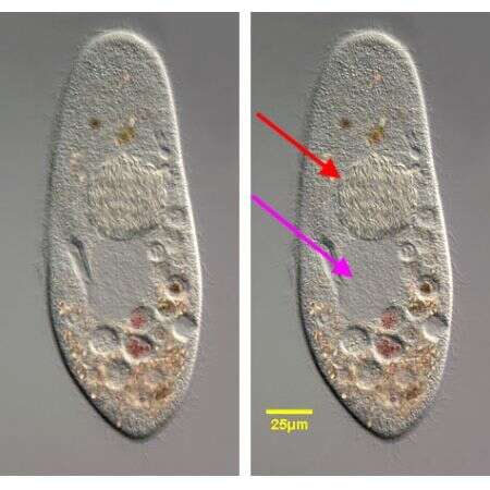
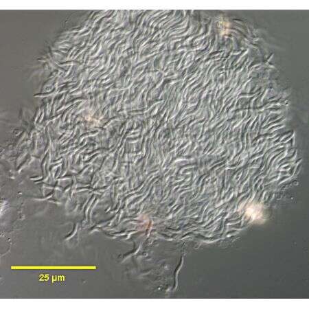
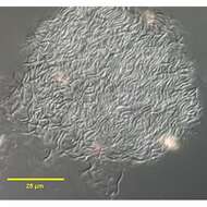
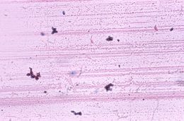
A mass of bacterial endosymbiont Holospora undulata (ex Hafkine, 1890) Gromov and Ossipov, 1981occupying the micronucleus of Paramecium caudatum (Ehrenberg,1833).DIC. A detailed discussion of the genus Holospora is available at http://141.150.157.117:8080/prokPUB/chaprender/jsp/showchap.jsp?chapnum=355&initsec=01_00
-
-
A mass of bacterial endosymbiont Holospora undulata (ex Hafkine, 1890) Gromov and Ossipov, 1981occupying the micronucleus of Paramecium caudatum (Ehrenberg,1833).DIC. A detailed discussion of the genus Holospora is available at http://141.150.157.117:8080/prokPUB/chaprender/jsp/showchap.jsp?chapnum=355&initsec=01_00
-
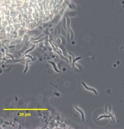
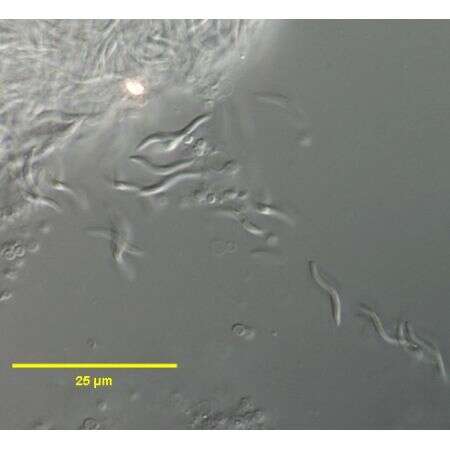
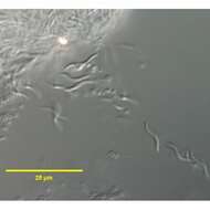
These cells of the endosymbiont Holospora undulata (ex Hafkine, 1890) Gromov and Ossipov, 1981are escaping from the micronucleus of a ruptured cell of the host, Paramecium caudatum (Ehrenberg,1833). Phase contrast.
-
These cells of the endosymbiont Holospora undulata (ex Hafkine, 1890) Gromov and Ossipov,1981are escaping from the micronucleus of a ruptured cell of the host, Paramecium caudatum (Ehrenberg,1833).
-
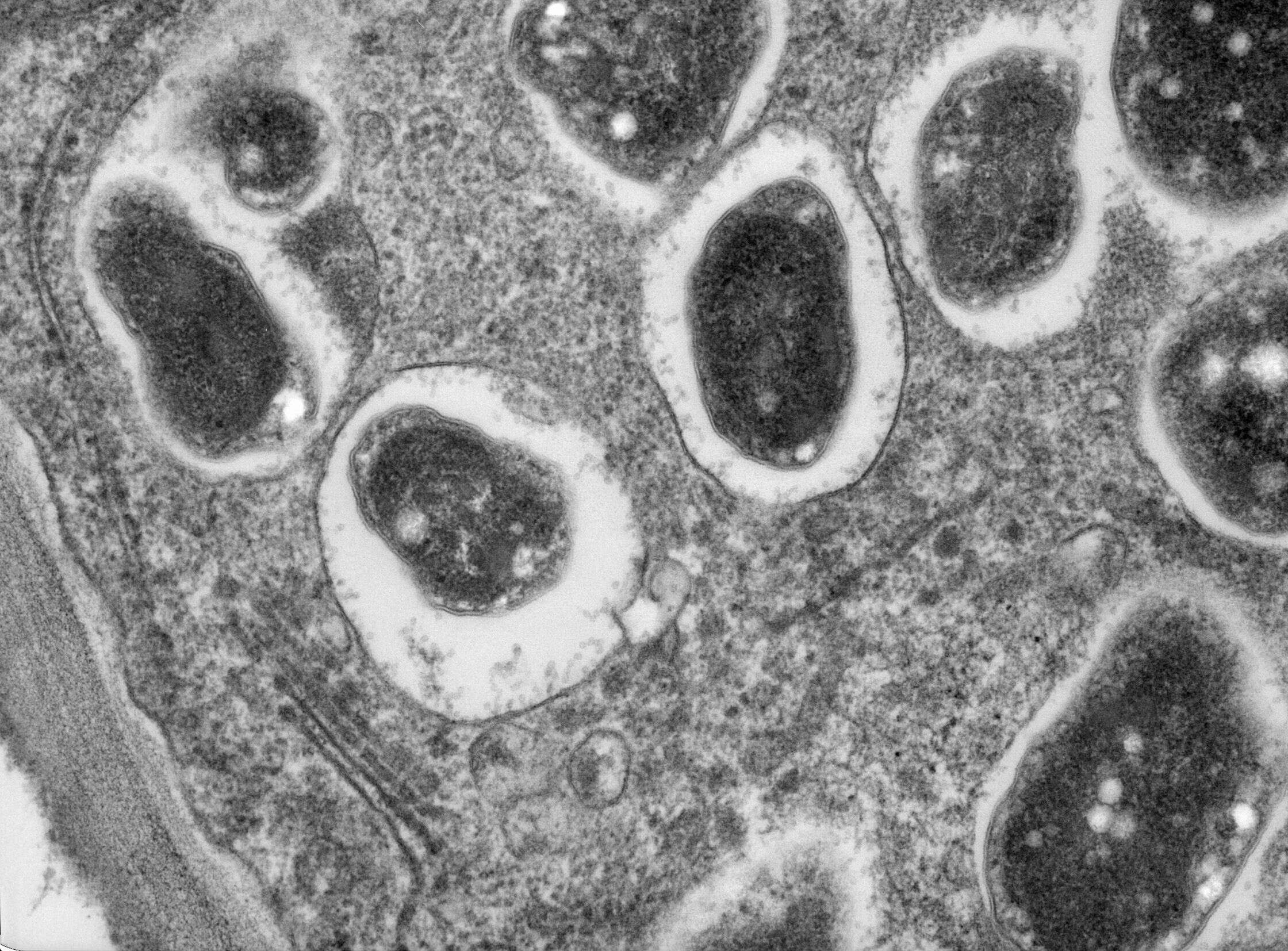
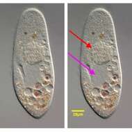
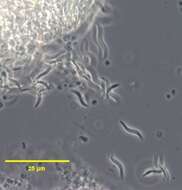
This is a detail of the grossly swollen micronucleus of a ruptured Paramecium caudatum (Ehrenberg,1833) infected with Holospora undulata (ex Hafkine, 1890) Gromov and Ossipov. The micronucleus has burst releasing individual cells of H. undulata..DIC.
-
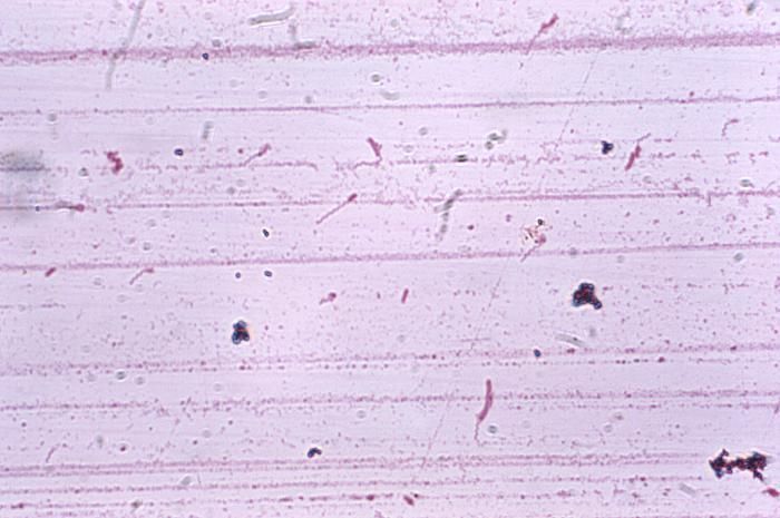
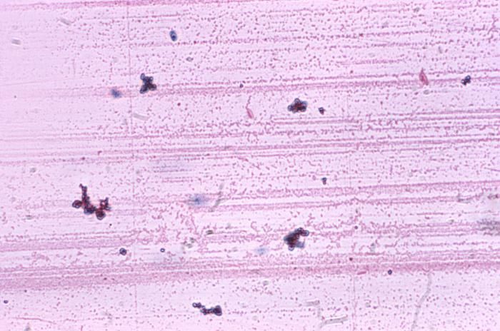
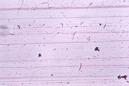
Magnified 1000X, this Liefsons flagella stained photomicrograph revealed the presence of a number of flagellated Brevundimonas diminuta, formerly known as Pseudomonas diminuta.Created: 1975
-
Magnified 1000X, this Liefsons flagella stained photomicrograph revealed the presence of a number of flagellated Brevundimonas diminuta, formerly known as Pseudomonas diminuta.Created: 1975
-
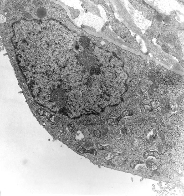
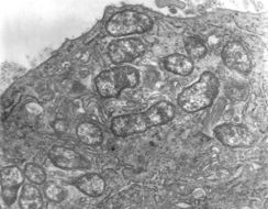
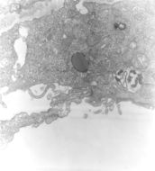
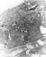
This 1976 transmission electron micrograph (TEM) depicted a hypertrophic peritoneal mesothelial cell of mouse that had been experimentally infected intraperitoneally with Orientia tsutsugamushi rickettsial micro-organisms. In this photomicrograph there were several organisms visible free within the mesothelial cell's cytoplasm.Created: 1976
-
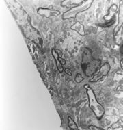
This 1978 transmission electron micrograph (TEM) depicted a brain capillary of a mouse that had been experimentally infected intravenously with Orientia tsutsugamushi rickettsial micro-organisms. Revealed by this TEM, was the presence of pericapillary hemorrhage and edema. Several of the O. tsutsugamushi organisms were visible within the cytoplasm of a degenerating capillary endothelial cell.Created: 1978
-
This 1978 transmission electron micrograph (TEM) depicted a brain capillary of a mouse that had been experimentally infected intravenously with Orientia tsutsugamushi rickettsial micro-organisms. This photomicrograph revealed three O. tsutsugamushi organisms visible within the cytoplasm of the host endothelial cell.Created: 1978
-
This 1978 transmission electron micrograph (TEM) depicted a brain capillary of a mouse that had been experimentally infected intravenously with Orientia tsutsugamushi rickettsial micro-organisms. This TEM revealed that one organism was budding from the luminal surface of a hypertrophic capillary endothelial cell, still covered by a third layer consisting of the host cell's plasma membrane. Others are visible free within the endothelial cell's cytoplasm.Created: 1978
-
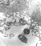
This 1976 transmission electron micrograph (TEM) depicted a hypertrophic peritoneal mesothelial cell of mouse that had been experimentally infected intraperitoneally with Orientia tsutsugamushi rickettsial micro-organisms. In this TEM, several organisms were visible, free within the host cell's cytoplasm. One O. tsutsugamushi appeared within a phagocytic vacuole, still bearing a third outer membrane layer of probable host cell origin.Created: 1976
-
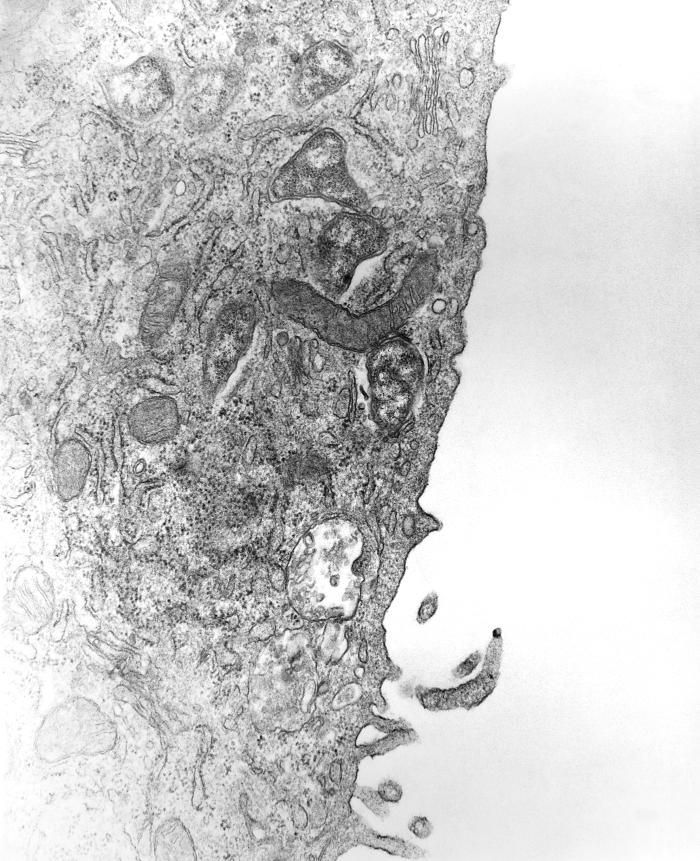
This 1976 transmission electron micrograph (TEM) depicted a hypertrophic peritoneal mesothelial cell of mouse that had been experimentally infected intraperitoneally with Orientia tsutsugamushi rickettsial micro-organisms. A disintegrating organism was shown within a host cell's phagocytic vacuole.Created: 1976
-
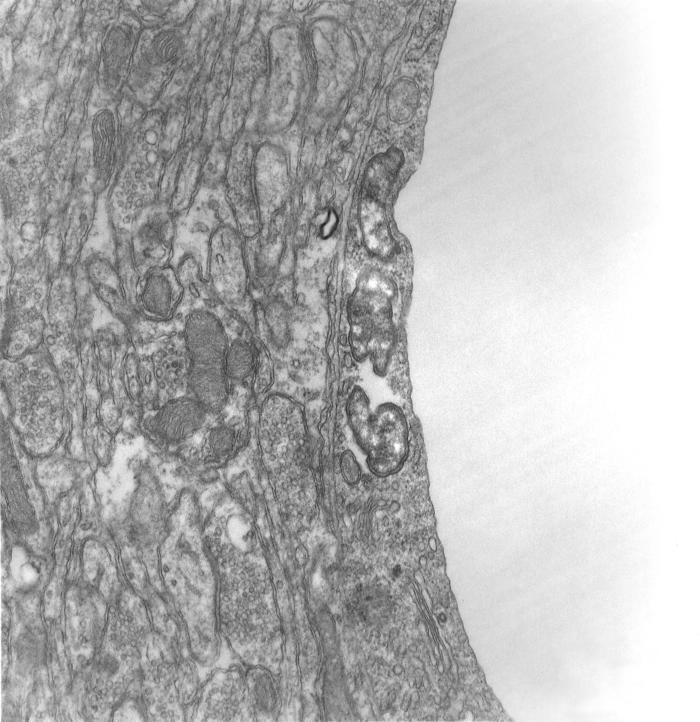
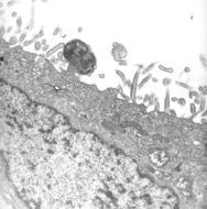
This 1976 transmission electron micrograph (TEM) depicted a peritoneal mesothelial cell from a mouse that had been experimentally infected intraperitoneally with Orientia tsutsugamushi rickettsial micro-organisms. Captured here in this TEM was the initiation of the process of phagocytosis being carried out on one of the O. tsutsugamushi micro-organisms.Created: 1976
-
This 1976 transmission electron micrograph (TEM) depicted peritoneal mesothelial cells from a mouse that had been experimentally infected intraperitoneally with Orientia tsutsugamushi rickettsial micro-organisms. This TEM revealed that the extracellular organisms were covered with a distinct third outer membrane of probable host cell origin.Created: 1976
-
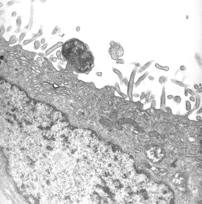
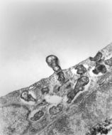
This 1976 transmission electron micrograph (TEM) depicted a hypertrophic peritoneal mesothelial cell of mouse that had been experimentally infected intraperitoneally with Orientia tsutsugamushi rickettsial micro-organisms. This micrograph showed one organism as it was in the process of budding from the luminal cell surface, still covered by a third layer, consisting of the host cell's plasma membrane. Others are visible free within the host cell's cytoplasm.Created: 1976
-
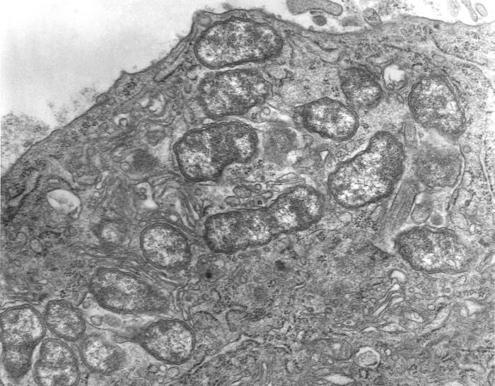
This 1976 transmission electron micrograph (TEM) depicted a hypertrophic peritoneal mesothelial cell of mouse that had been experimentally infected intraperitoneally with Orientia tsutsugamushi rickettsial micro-organisms. This particular photomicrograph revealed that there were multiple organisms free within the host cell's cytoplasm.Created: 1978
-
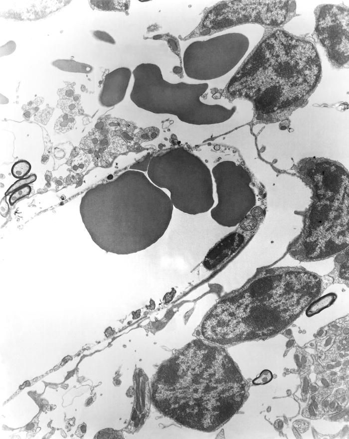
This 1978 transmission electron micrograph (TEM) depicted a brain capillary of mouse that had been experimentally infected intravenously with Orientia tsutsugamushi rickettsial micro-organisms. This photomicrograph revealed that the capillary lumen was partially occluded by a thrombus, and a hypertrophic, degenerating, apparently detached endothelial cell containing several visible organisms free within its cytoplasm.Created: 1978
-
This 1978 transmission electron micrograph (TEM) depicted a brain capillary of mouse that had been experimentally infected intravenously with Orientia tsutsugamushi rickettsial micro-organisms. In this micrographic view revealed pericapillary edema, the capillary lumen was partially occluded by a thrombus, and flanking the thrombus, a hypertrophic endothelial cell containing a visible organism free within its cytoplasm.Created: 1978
-
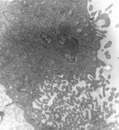
This 1978 transmission electron micrograph (TEM) depicted a brain capillary of mouse that had been experimentally infected intravenously with Orientia tsutsugamushi rickettsial micro-organisms. In this micrographic view, a host cell endothelial cell was shown to contain many organisms free within its cytoplasm.Created: 1978
-
This 1976 transmission electron micrograph (TEM) depicted a hypertrophic peritoneal mesothelial cell of mouse that had been experimentally infected intravenously with Orientia tsutsugamushi rickettsial micro-organisms. This micrograph captured a dividing organism while it was visible free within the host cell's cytoplasm.Created: 1976
-
This 1976 transmission electron micrograph (TEM) depicted a hypertrophic peritoneal mesothelial cell from mouse that had been experimentally infected intraperitoneally with Orientia tsutsugamushi rickettsial micro-organisms. This micrograph captured an organism as it appeared within a phagocytic vacuole. An outer third membrane, of probable host cell origin, was disintegrating, leaving behind only its electron-dense remnants.Created: 1976


