Depth range
provided by World Register of Marine Species
305-341 m.
Zhadan, Anna; Atroshchenko, Margarita. (2012). A new species of Fauveliopsidae (Annelida) from the North Sea. ZooKeys. 181: 1-10.
- license
- cc-by-4.0
- copyright
- WoRMS Editorial Board
Distribution
provided by World Register of Marine Species
NE Atlantic Ocean: northeast part of the North Sea (off Norway).
Zhadan, Anna; Atroshchenko, Margarita. (2012). A new species of Fauveliopsidae (Annelida) from the North Sea. ZooKeys. 181: 1-10.
- license
- cc-by-4.0
- copyright
- WoRMS Editorial Board
Habitat
provided by World Register of Marine Species
Muddy sediment with small admixture (0.8-7.7%) of fine and medium sand, at shelf depths (about 300-350 m).
Zhadan, Anna; Atroshchenko, Margarita. (2012). A new species of Fauveliopsidae (Annelida) from the North Sea. ZooKeys. 181: 1-10.
- license
- cc-by-4.0
- copyright
- WoRMS Editorial Board
Description
provided by Zookeys
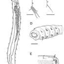
Adult specimens 6–8 mm long and 0.35–0.4 mm wide with 22 chaetigers. Only one specimen has 23 chaetigers. Body slender, often c–shaped or s-shaped by fixation, colorless in preserved material. Four anteriormost chaetigers swollen, following segments cylindrical, without clear borders (Fig. 2A, 3A, B).
Cuticle thick (about 8 μm), smooth, with very thin ring wrinkles, bearing scattered minute inconspicuous micropapillae (Fig. 3D, 4C), visible under higher magnification and in SEM. Relatively larger micropapillae surrounding inverted parts – prostomium and pygidium (Fig. 4B).
Ventral nerve cord visible by transparency. Ganglia longitudinally elongated and indistinctly separated from each other.
Prostomium small, from round to triangular, lacking appendages, with ciliary nuchal organs, without eyes (Fig. 2D, 3B). In most specimens prostomium completely retracted into peristomium (Fig. 4A). Peristomium forms complete ring; it bears micropapillae.
Parapodia biramous, best developed on chaetigers 1–4 and hardly distinct on posteriormost segments except last one (22). Interramal papillae short–stalked, pyriform, situated midway between noto- and neuropodia (Fig. 3C, 4A, B, C, E, F). Two epidermal glands visible in each parapodium.
Chaetigers 1–4 bear thick sigmoidal hirsute chaetae. Two chaetae in notopodia and four (2 long and 2 shorter ones) in neuropodia (Fig. 2B, 4C). Some specimens possess five instead of four neurochaetae in anterior segments. Neurochaetae mostly bidentate (Fig. 4D), notochaetae usually unidentate, sometimes slightly bidentate. Some specimens possess 3–4 bidentate neurochaetae whereas others have only 1–2 bidentate neurochaetae. Both short and long neurochaetae can be bidentate or unidentate. Two specimens have transitional fifth segment with chaetae more similar to acicular chaetae of anterior four segments than to thin capillary chaetae of rest of the body.
Chaetigers 5–21 bear one thin capillary chaeta and one accessory very short, thin and barely distinct capillary chaeta per ramus (Fig. 2C, 4E, F). Notopodial chaetae thinner and shorter than neuropodial ones; accessory chaetae in notopodia often absent.
Chaetiger 22 bears one thick acicular chaeta in notopodia and three capillary chaetae in neuropodia (Fig. 2E, 3C, 4B).
Paired genital papillae (Fig. 3E, 4F) situated at posterior edge of chaetiger 8 in all studied individuals with gametes as well as in specimens without recognizable gametes, except for one specimen with unpaired genital papilla.
Pygidium retracted within last chaetiger, anus terminal. Boundary between distal part of last segment and pygidium indistinct. Inverted part (distal to chaetae) with a number of conical papillae, which are larger than in rest of body (Fig. 2E, 3C, 4B).
Oocytes up to 90 μm in diameter, arranged in two ovisacs, extend through chaetigers 6–15, each contains 20–40 oocytes (Fig. 2A, 3A, E).
Sperm cells hardly visible through body wall, but observed in SEM in body cavity of dissected specimen. They have rounded heads about 3 μm in diameter, an acrosome with elongated distal part and free flagellum at least 10 μm long (Fig. 4G, H).
- license
- cc-by-3.0
- copyright
- Anna Zhadan, Margarita Atroshchenko
- bibliographic citation
- Zhadan A, Atroshchenko M (2012) A new species of Fauveliopsidae (Annelida) from the North Sea ZooKeys 181: 1–10
- author
- Anna Zhadan
- author
- Margarita Atroshchenko
Distribution
provided by Zookeys
Northeast part of the North Sea.
- license
- cc-by-3.0
- copyright
- Anna Zhadan, Margarita Atroshchenko
- bibliographic citation
- Zhadan A, Atroshchenko M (2012) A new species of Fauveliopsidae (Annelida) from the North Sea ZooKeys 181: 1–10
- author
- Anna Zhadan
- author
- Margarita Atroshchenko