-
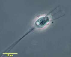
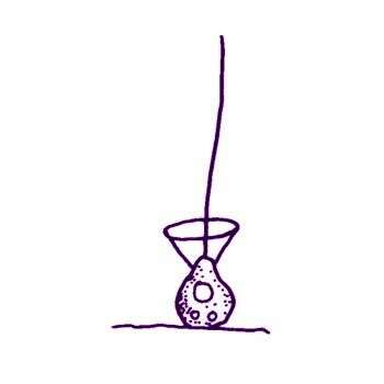
Salpingoeca huxleyi Ellis, 1930. Cell globular. Body of lorica egg-shaped, with a wide, sturdy neck which bulges towards its own base. This neck appears to be marked off, at its actual base, from the lorica proper by a costa-like ring. This ring is probably an optical effect due to the lorica neck being set in a shallow depression or in-fold of the lorica body. A further peculiarity is that the neck of the cell is never extended beyond the distal limit of the lorica neck, so that the base of the collar is always within its everted rim. Collar and flagellum normal. Nucleus conspicuous. Contractile vacuoles: two. Peduncle length variable. Length of body of lorica: 8 microns Length of neck of loriea: 4 microns Width at centre of neck of lorica: 3 microns Greatest width of lorica: 6.5 microns Peduncle length: 16-32 microns
-
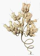
Codonocladium cymosum (Kent, 1880) Boucaud-Camou, 1967. Cells symmetrically ovate, located on short independent stalks, at the ends of a branching stalk-system, cells 5 microns long, main stalks 51-102 microns
-
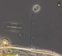
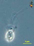
This phase contrast image of cell, no flagellum, and lorica taken from ATCC strain 50455 in April 2006.
ATCC data on this organism.
-
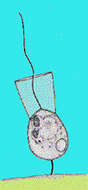
Portrait of Diploeca flava a salpingoecid choanoflagellate. The cell body (protoplast) Is seen within a double-walled lorica. Iron salts color the thick outer layer brown. The clear, thin, trumpet-shaped inner layer of the lorica is seen projecting beyond the thick outer layer. The collar of long microvilli surrounding the single terminal flagellum is seen extending from the protoplast beyond the inner neck of the lorica. A single contractile vacuole is seen in the protoplast. The flagellum creates water currents directing food particles to the microvilli. Food particles are ingested at the bases of the microvilli. This genus is distinguished from Salpingoeca by its double-walled lorica. Diploeca may be confused with Diplosigopsis. In the latter genus what appears to be a clear trumpet-like extension of the lorica is actually a cytoplasmic collar extending from the protoplast to surround the wreath of microvilli (for an excellent discussion and illustration of the differences between these two genera see P. Bourrelly (1981) Les Algues D'eau Douce (Tome II) pp 130-135. Societe des Editions Boubee. Collected from a freshwater pond near Boise, Idaho December 2003. DIC optics.
-
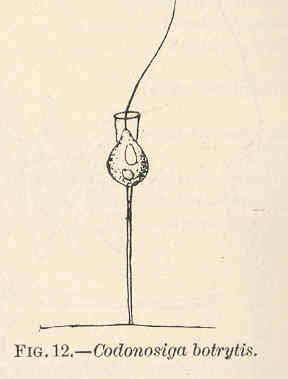
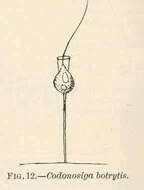
Codonosiga botrytis.
-
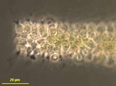
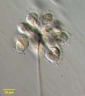
Desmarella moniliformis Kent, 1880. Cells 6.4 microns, symmetrically ovoid, arranged in single chain-like series, each colony containing from two to as many as eight individuals, nucleus spherical, subcentral, two or more contractile vacuoles, posteriorly located.
-
Codonocladium candelabrum.
-
Codosiga (co-dough-sigh-ga) also known as Codonosiga (co-dough-know-sigh-ga), choano-flagellate (collar flagellate) with a single apical flagellum surrounded by a collar of fine pseudopodia. This cell is stressed and has become spherical and the collar has been withdrawn. Normally many cells are attached to the stalk. Phase contrast.
-
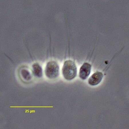
In vivo portrait of the colonial choanoflagellate, Desmarella moniliformis (Kent,1878). Ovoid cells are joined at lateral surfaces foming linear or slightly crescentic colonies.Cells have single anterior apical flagellum surronded by a collar of microvilli.Cells lack a pedicel.Collected near Boise, Idaho (43°38'21.10"N 116°11'10.78"W elev. 2908 ft.) from an ice-covered temporary puddle containing leaf litter and dead grass.November, 2005.Phase contrast.
-
Codosiga (co-dough-sigh-ga) also known as Codonosiga (co-dough-know-sigh-ga),choano-flagellate (collar flagellate) with a single apical flagellum surrounded by a collar of fine pseudopodia. Normally many cells are attached to the stalk. Phase contrast.
-
In vivo portrait of the colonial choanoflagellate, Desmarella moniliformis (Kent,1878). Ovoid cells are joined at lateral surfaces foming linear or slightly crescentic colonies.Cells have single anterior apical flagellum surronded by a collar of microvilli.Cells lack a pedicel.Collected near Boise, Idaho (43°38'21.10"N 116°11'10.78"W elev. 2908 ft.) from an ice-covered temporary puddle containing leaf litter and dead grass.November, 2005.DIC.
-
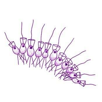
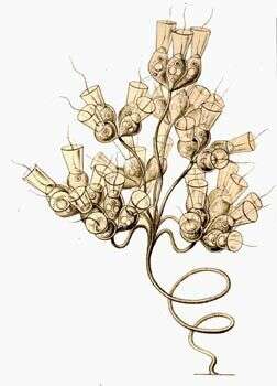
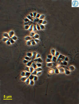
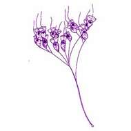
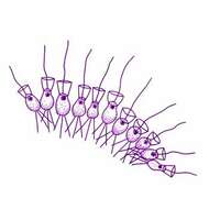
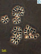
Codosiga, a collar flagellate. This genus includes solitary and colonial forms. Here we see several colonies of cells forming tight aggregates at the end of a long stiff stalk. Each cell has a single apical flagellum surrounded by a collar of fine pseudopodia. The collar captures food swept against it by the action of the flagella. Phase contrast.
-
Monosiga (mono-sigh-ga) a naked choanoflagellate (collar-flagellate). The cell body is small - about 5 microns. A single flagellum projects from the anterior end. It is surrounded by a collar of fine pseudopodia. Beating of the flagellum draws a current of water through the collar and particles of food are trapped against the collar. Pseudopodia can then enclose the food so that it can be digested. Animations by Rosemary Arbur of flagellar beat patterns are available
here. Phase contrast.
-
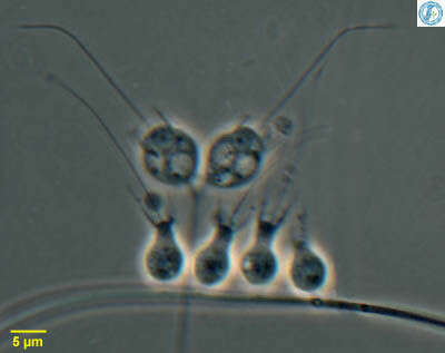
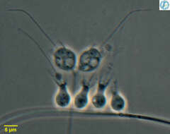
Codosiga - collar flagellate, two cells attached to a common stalk with a collection of Salpingoeca - also a collar flagellate. All these cells have a single apical flagellum that is surrounded with a collar of fine pseudopodia that appears as two dark lines, one to either side of the flagellum in this micrograph. Feed on suspended bacteria. From Lake Donghu, China Phase contrast micrograph.
-
Monosiga (mono-sigh-ga) a naked choanoflagellate (collar-flagellate). The cell body is small - about 5 microns A single flagellum projects from the anterior end. It is surrounded by a collar of fine pseudopodia. Beating of the flagellum draws a current of water through the collar and particles of food are trapped against the collar. Pseudopodia can then enclose the food so that it can be digested. Animations by Rosemary Arbur of flagellar beat patterns are available
here. Phase contrast.
-
-
-
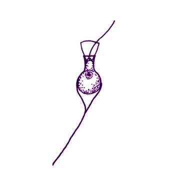
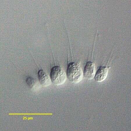
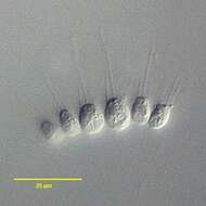
Codosiga botrytis (Ehrenberg) Kent, 1880. Cells are 3-5 x 5-15 microns long, cells are ovoid or spherical with posterior thin stalk. The cell body, and a small part of the stalk, is enclosed in a thin lorica which is barely visible in the light microscope. Two or more cells may be attached by short individual stalks to a common stalk. The cells may break away from the stalk and swim freely, in this state numerous short thin pseudopodia radiate from the cell posterior.
-
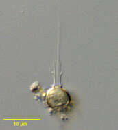
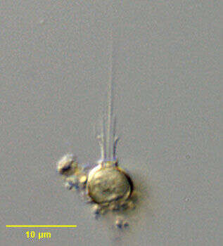
Monosiga (mon-owe-sigh-ga), a choanoflagellate (collar flagellate), here many cells have colonised a small piece of detritus. Each globular cell has a conical collar around the single apical flagellum. Phase contrast.
-
Codosiga botrytis.
-
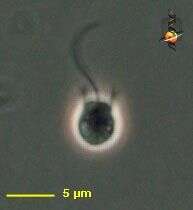
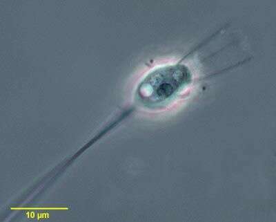
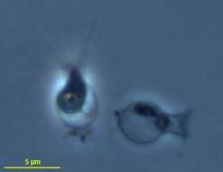
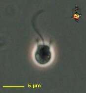
Portrait of Monosiga, a choanoflagellate without a visible lorica. A collar of pseudopodia which trap food particles surrounds a central flagellum. The particles are transported to the base of the collar where they are ingested. This is an unusually large specimen. The cell body is usually about 5 microns in length. The nucleus, food vacuoles and contractile vacuole are well seen here. This species is stalked while others are not, the cell body attaching directly to the substrate. From a freshwater pond near Boise, Idaho. Phase contrast.
-
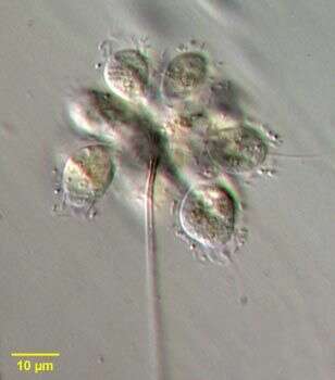
Portrait of choanoflagellate, Codosiga botrytis. Cells are clustered on simple, terminally branching stalk. Circumferential collar of microvilli can be seen. Single long flagellum directs food particles to the outside surface of the microvilli along which they move to be ingested at the apex of the cell. Adhering food particles are seen in these images. From a freshwater pond near Boise, Idaho. Phase contrast.
-
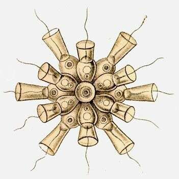
Monosiga ovata Kent, 1880. The ovoid cell is approximately 4 microns long or 15 microns when measured from the tip of the flagellum to the distal end of the stalk. At the anterior end of the cell a ring of 20-25 tentacles, almost equal in length, forms a collar which encircles the single flagellum. The latter projects beyond the tentacles and terminates in a conspicuous hair point. The cell body and the base of the tentacles are closely invested by a delicate membranous sheath and this tapers posteriorly to form the stalk or peduncle which attaches the cell to the substratum. The apparent looseness of the sheath in shadowcast whole mounts is probably caused by the shrinkage of the protoplast during drying. Transverse sections of the cell in the region of the tentacles and cell body show that the sheath fits closely although it is ridged at intervals. That part forming the stalk sometimes has a fibrillar appearance but it is not clear whether this is a partial disintegration caused by drying or a genuine arrangement for attaching the cell to the substratum. In section the sheath is seen to be composed of two regularly spaced layers, the surfaces of which are covered with a fine fibrillar deposit possibly of mucilage.
-
Portrait of choanoflagellate, Codosiga botrytis. Cells are clustered on simple, terminally branching stalk. Circumferential collar of microvilli can be seen. Single long flagellum directs food particles to the outside surface of the microvilli along which they move to be ingested at the apex of the cell. Adherent food particles are seen in these images. From a freshwater pond near Boise, Idaho. Oblique illumination.