-
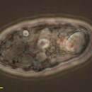
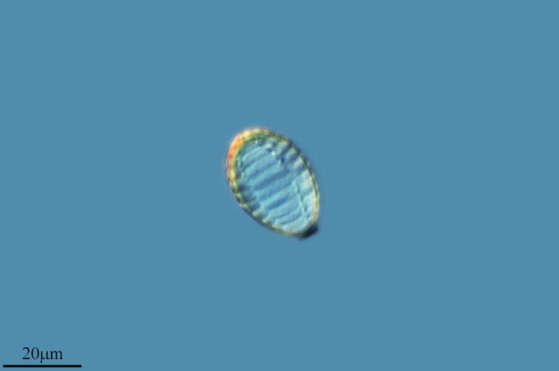
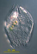
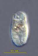
Protaspis obliqua Skuja, 1939. Cells are slightly oval or roundish, 8 to 32 microns long, 10 to 27 microns wide, dorso-ventrally flattened and with thickened cortex. There is a ventral median groove, cell indented anteriorly and posteriorly where the groove meets margin. Subapically, the right margin of the groove forms a protrusion. With two flagella inserting under the protrusion, the anterior flagellum is about 0.5 times the length of the cell and the posterior flagellum is about 0.5 to 1.5 times the length of the cell. The nucleus is without nuclear caps, is located subapically in a median position, is rounded and is 5 to 13 microns in diameter. The cells may contain food particles or diatom up to 24 microns long.
-
Galende, Castile and Len, Spain
-
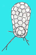
Members of testate amoebae group Euglyphidae form siliceous plates for constructing shell. Sample collection by Martin Kreutz from Simmelried near Konstanz(Baden-Wuerttemberg, Germany). This image was taken using Zeiss Universal with Olympus C7070 CCD camera.
-
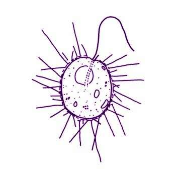
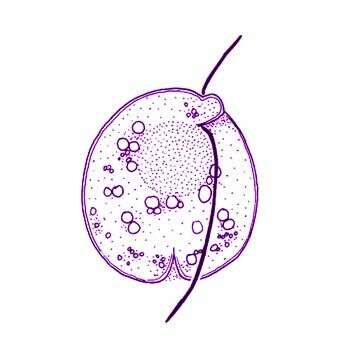
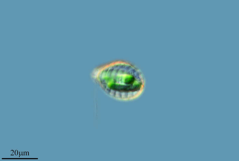
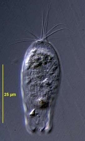
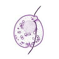
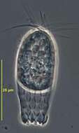
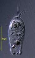
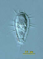
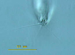
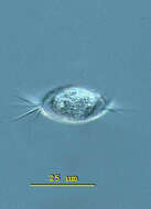
Thaumatomastix salina (Birch-Andersen) Beech and Moestrup, 1986. Cells are ovoid (7-12 microns x 8-15 microns), slightly compressed dorso-ventrally, and have a long flagellum 3/4-5/4 of the cell length. A short flagellum, which is rarely visible, emerges together with the long flagellum from what appears to be a very slight groove or depression located latero-anteriorly. A furrow-like structure is often noted running from the flagellar bases to the cell midline. Cells are solitary, and are most often observed attached to pieces of detritus. Cells occasionally move in a creeping motion, with the long flagellum trailing and gliding over the coverslip. In some cases cells swim freely with the long flagellum making irregular, arhythmical flicking motions. The cell cytoplasm has a granular appearance and is devoid of any kind of chloroplast, a diffuse area of a pale orange colour can often be noticed in the central part of the cell when phase contrast oil immersion optics are used. One cell was noted in an early stage of division where both flagella had replicated. Spine scales, varying in length, radiate from the entire cell surface. Flattened cells slough off their scales and scales of a second type, spineless body scales, can then be seen to be elliptical in outline.
-
Galende, Castille and Leon, Spain
-
Apertural plates of Euglypha cristata Leidy, 1874. Found in a soil sample from Pyhä-Luosto National Park, Finland. DIC.
-
Ribadelago, Castille and Leon, Spain
-
Euglypha (you-gligh-fah) cristata has an elongate shell that is composed of siliceous scales. The point of the shell shows a characteristic tuft of three to eight spines. The circular aperture is bordered by a single row of five to six denticulate scales. Pseudopodia are rarely extended. Differential interference contrast.
-
Ribadelago de Franco, Castille and Leon, Spain
-
Euglypha cristata Leidy, 1874. Found in a soil sample from Pyhä-Luosto National Park, Finland. Phase contrast.
-
Ribadelago de Franco, Castille and Leon, Spain
-
Euglypha cristata Leidy, 1874. Found in a soil sample from Pyhä-Luosto National Park, Finland. DIC.
-
Ribadelago, Castille and Leon, Spain
-
Euglypha (you-gligh-fah) filifera has an elongate shell that is composed of oval siliceous scales. The 15 micron long spines pointed out from the entire lateral edge of the shell. The nucleus is visible in the posterior third of the cell. Differential interference contrast.
-
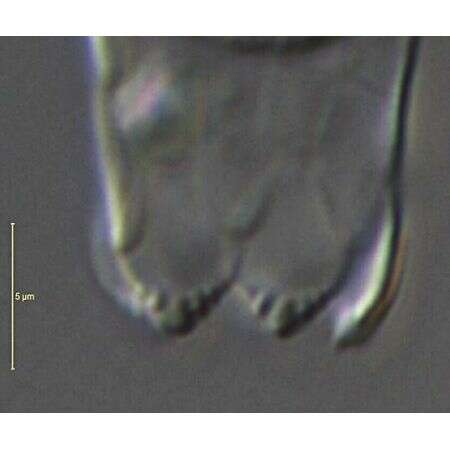
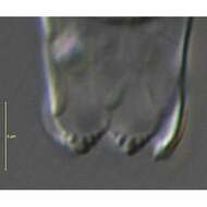
Euglypha (you-gligh-fah) filifera has an elongate shell that is composed of oval siliceous scales. The 15 micron long spines pointed out from the entire lateral edge of the shell. View of the aperture surrounded by denticulate scales. Long filopodia extended from the aperture. The aperture measures 6 microns in diameter. Differential interference contrast.
-
Euglypha (you-gligh-fah) filifera has an elongate shell that is composed of oval siliceous scales. The 15 micron long spines pointed out from the entire lateral edge of the shell. Image of Euglypha filifera lorica. The denticulate scales which border the circular aperture are visible. Each plate is measuring 6 X 8 microns on average. Differential interference contrast.
-
Euglypha (you-gligh-fah) filifera has an elongate shell that is composed of oval siliceous scales. The 15 micron long spines pointed out from the entire lateral edge of the shell. Apical view on the shell of Euglypha filifera. From this view it is evident that the shell is compressed and that the spines arise from the lateral edge of the shell. Differential interference contrast.
-
-
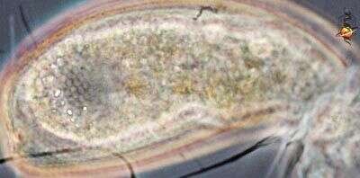
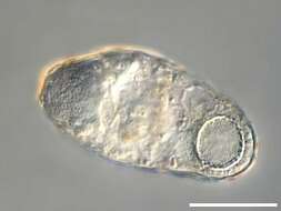
Trinema (try-knee-ma) enchelys, a testate amoeba. The focal plane on the circular aperture of the shell of Trinema enchelys. The aperture is located on the ventral surface and is surrounded by a number of rows of very minute scales. The central located contractile vacuole is visible and at the posterior end of the cell the macronucleus. This specimen was collected in a freshwater pond near Konstanz, Germany. Differential interference contrast.
-
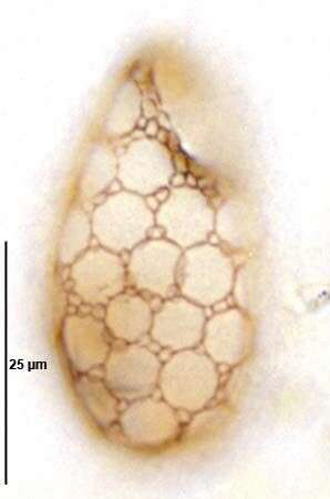
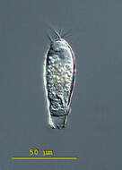
Trinema (try-knee-ma) enchelys, a testate amoeba. This specimen was collected in a freshwater pond near Konstanz, Germany. The focal plane is on the surface of the shell of Trinema enchelys. The shell is composed of transparent circular scales, each 5 - 6 mm in diameter. Differential interference contrast.
-
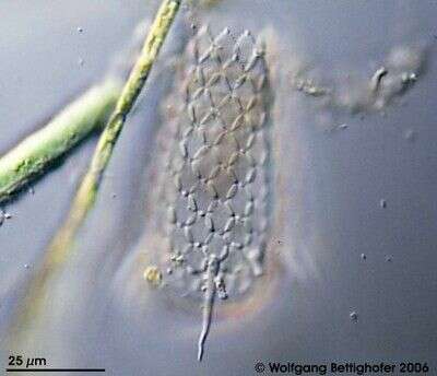
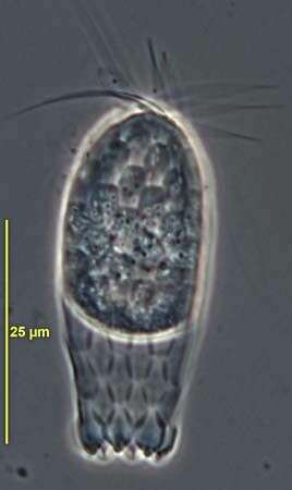
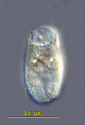
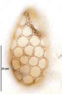
Trinema enchelys (EHRENBERG,1838) LEIDY,1878.Large incompletely overlapping circular shell plates with smaller peripherally placed oval shell plates. Collected from bottom sediments of an organically enriched freshwater pond near Boise, Idaho. December 2007. Protargol (see Foissner, W. Europ. J. Protistol., 27:313-330;1991).Brightfield.
-
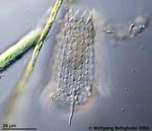
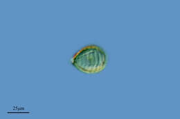
Scale bar indicates 25 µm. Sample from a wetland at the Pillersee (Tyrol, Austria). The image was built up using several photomicrographic frames with manual stacking technique. Images were taken using Zeiss Universal with Olympus C7070 CCD camera.Image under Creative Commons License V 3.0 (CC BY-NC-SA).
-
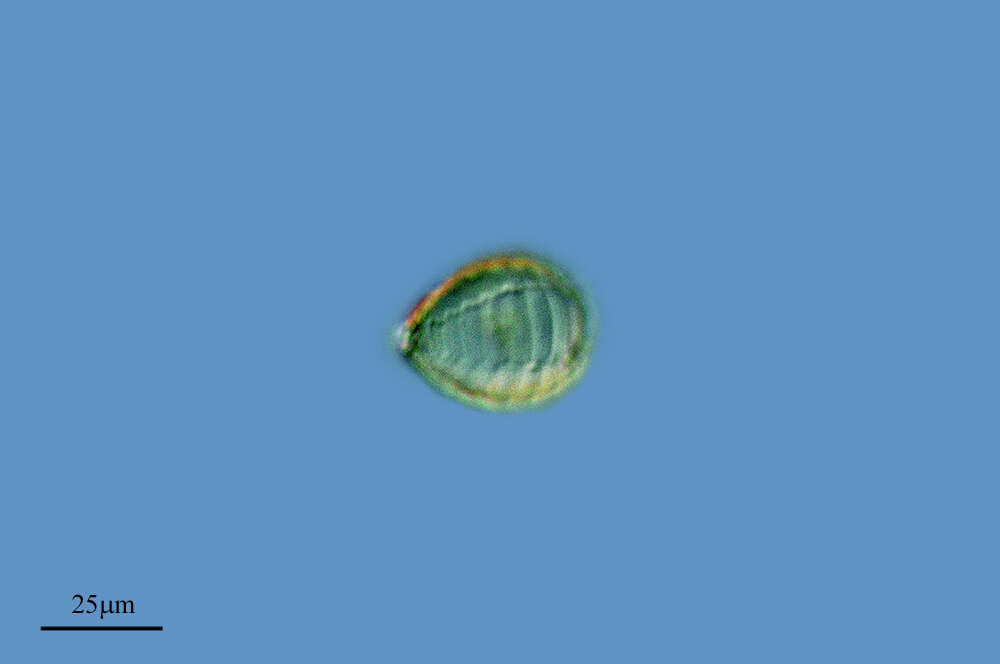
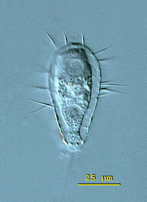
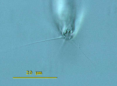
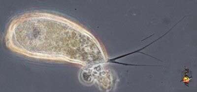
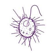
Cyphoderia (sigh-fo-dear-ee-a) is a shelled amoebae. This one has thin branching pseudopodia. The amoeba is protected by a loosely fitting lorica which has a single aperture through which the pseudopodia emerge. This form is thought to provide protection from the damaging effects of surface tension of water - something which many organisms living in the spaces between intertidal sand particles are likely to encounter as the tide goes out. Individual genera and species are mostly distinguished by the texture of the surface of the lorica. Not so common in marine habitats, more common in soils. Phase contrast.
-
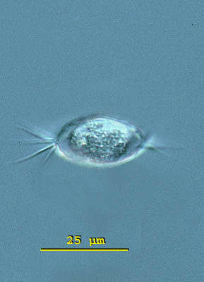
Cyphoderia (sigh-fo-dear-ee-a) is a shelled amoebae. This one has thin branching pseudopodia. The amoeba is protected by a loosely fitting lorica which has a single aperture through which the pseudopodia emerge. The lorica of has thin scales attached to the external surface. These are evident towards the left of the picture. Phase contrast.