-
S Pedro, Galicia, Spain
-
Galende, Castile and Len, Spain
-
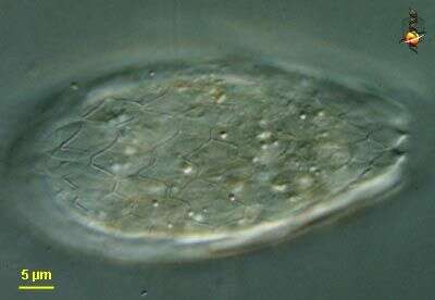
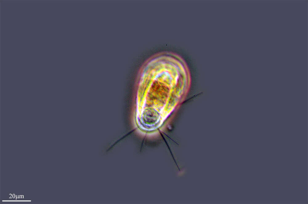
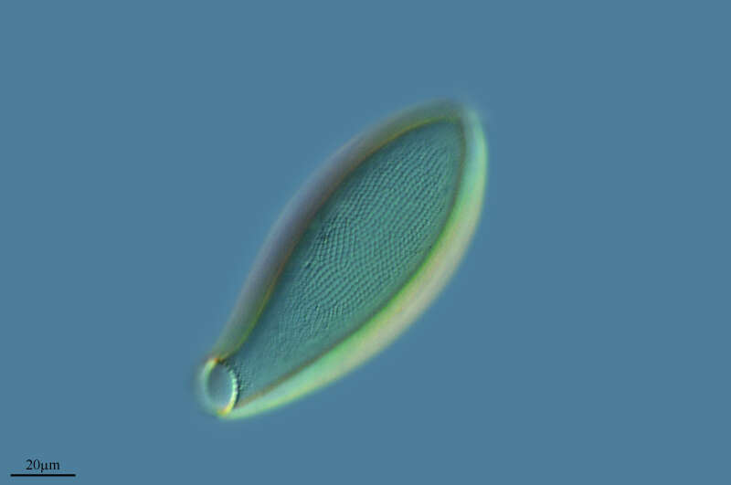
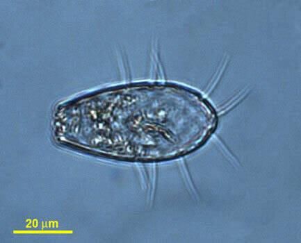
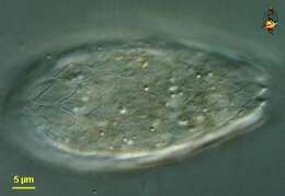
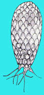
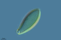
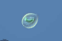
Euglypha (you-gly-fa) a shelled amoeba with filose pseudopodia (although this picture is only of the test) and with the test covered in small overlapping scales and a terminal aperture.Differential interference contrast.
-
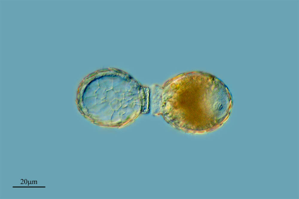
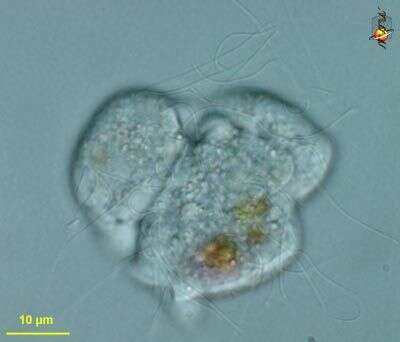
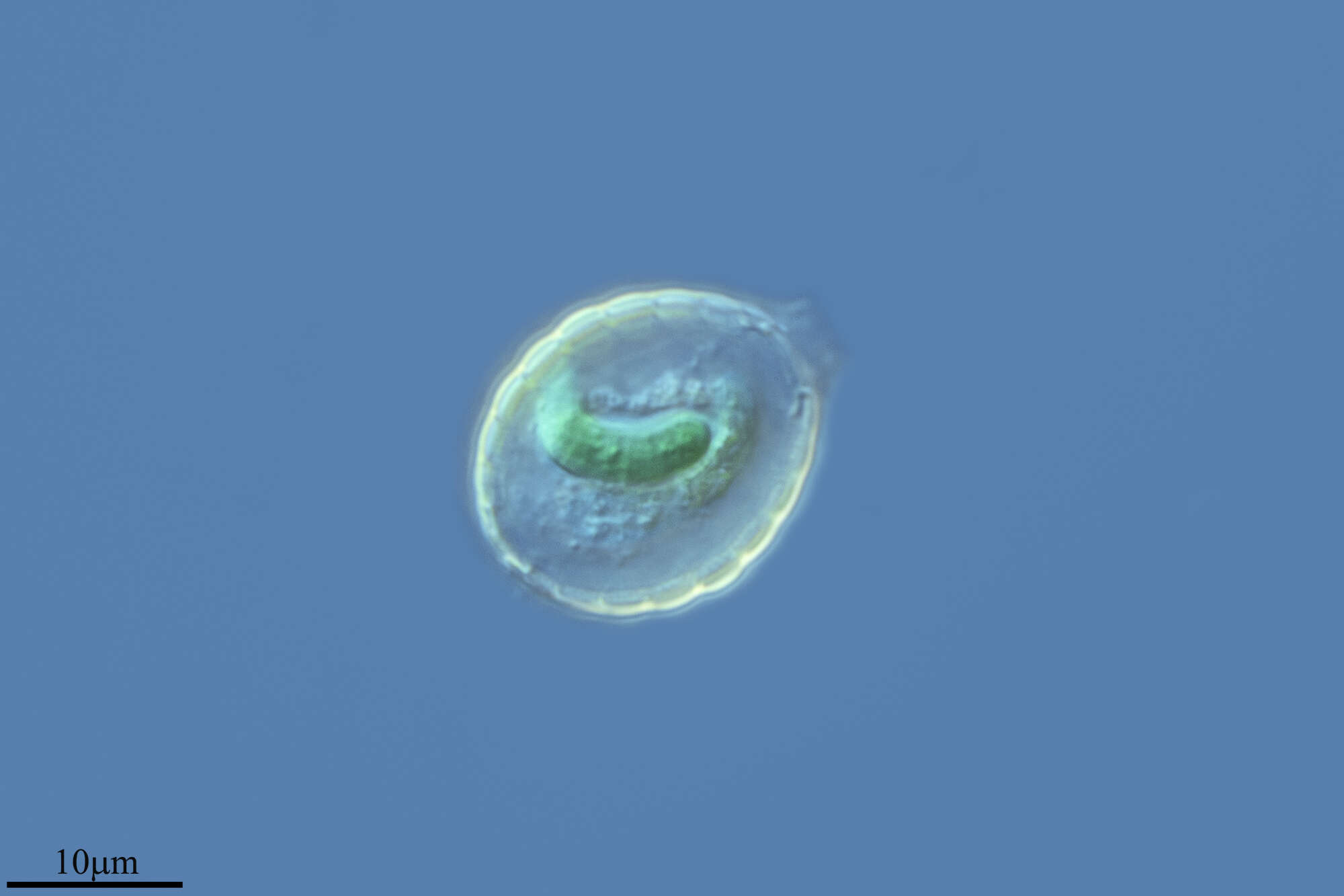
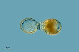
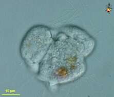
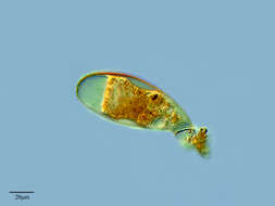
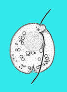
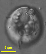
Protaspis (pro-tass-piss) is a medium-sized heterotrophic flagellate. Two flagella emerge close to each other from a point behind the apex of the cell and on the ventral side. The anterior flagellum is typically shorter than the posterior flagellum. The ventral side may give rise to pseudopodia which can enclose food - such as diatoms. In this image, many cells have fused into a syncitium. DIfferential interference microscopy.
-
San Martn de Castaeda, Castilla y Len, Espaa
-
Canencia, Madrid, Spain
-
Lumbreras, La Rioja, Spain
-
S Pedro, Galicia, Spain
-
Galende, Castile and Len, Spain
-
-
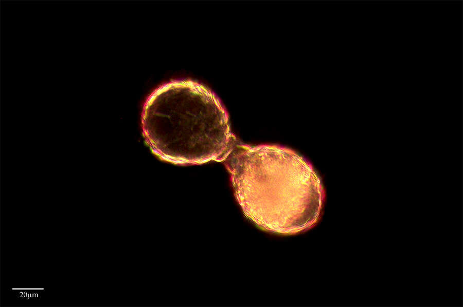
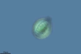
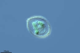
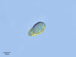
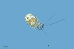
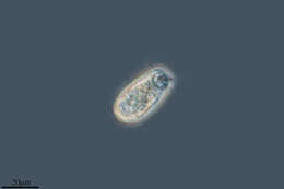
Biflagellate protist with a ventral furrow and anterior depression from where the two flagella emerge. Species not identified. Isolated by M. Virginia Sanchez Puerta from Sippewissett Pond, Woods Hole, MA, USA. Photographed using DIC microscopy.
-
Galende, Castille and Leon, Spain
-
Franceses, Canary Islands, Spain
-
Ribadelago de Franco, Castille and Leon, Spain
-
Cedar Swamp, Woods Hole, Massachusetts, USA. Photoed by Hwan Su Yoon.
-
-
Ribadelago, Castille and Leon, Spain
-
Matute, La Rioja, Spain
-
Galende, Castile and Len, Spain
-
Samples from Sediment at Cedar swamps, Woods Hole, Massachusatts. Photographed by Hwan Su Yoon.
-
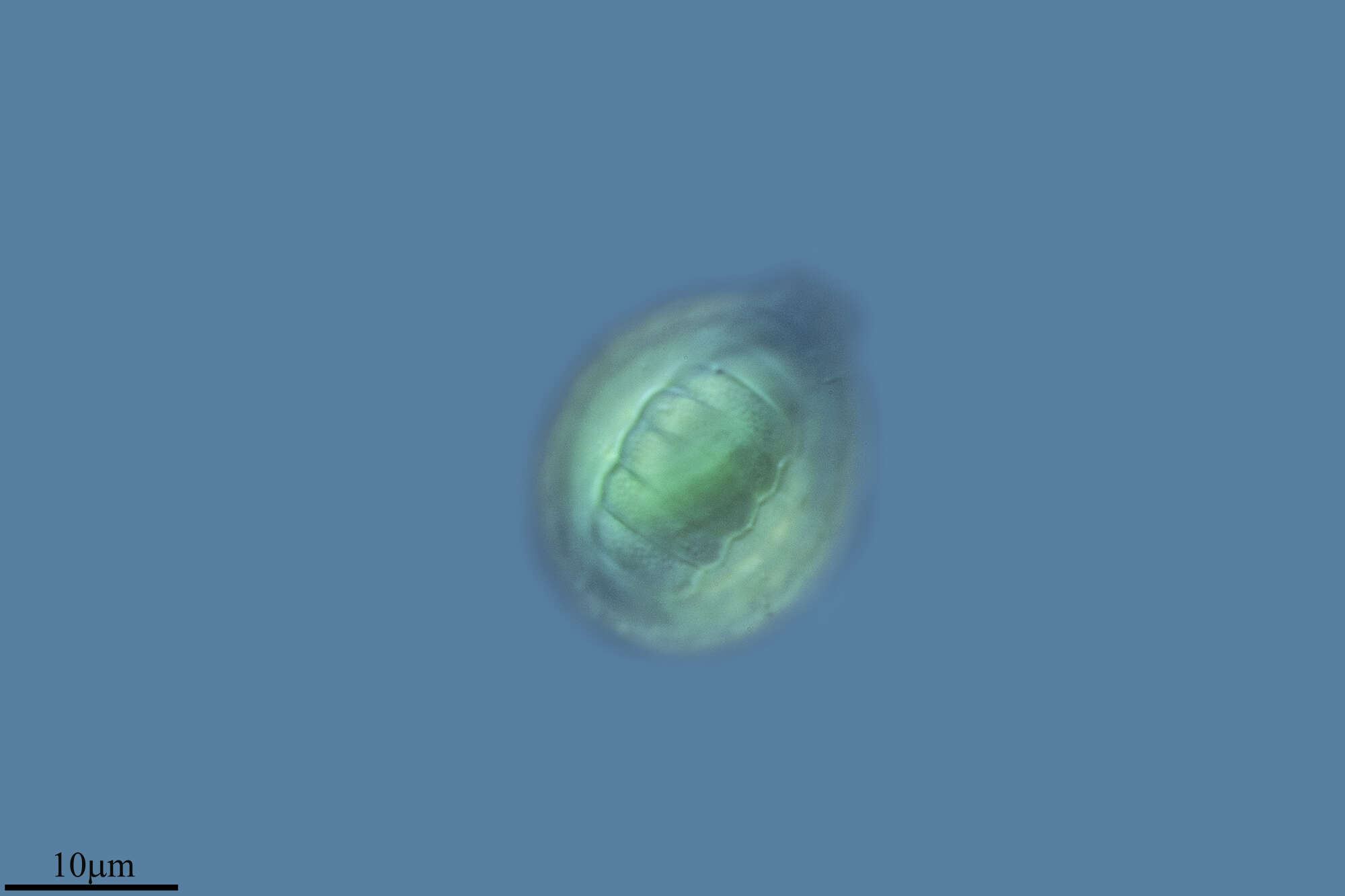
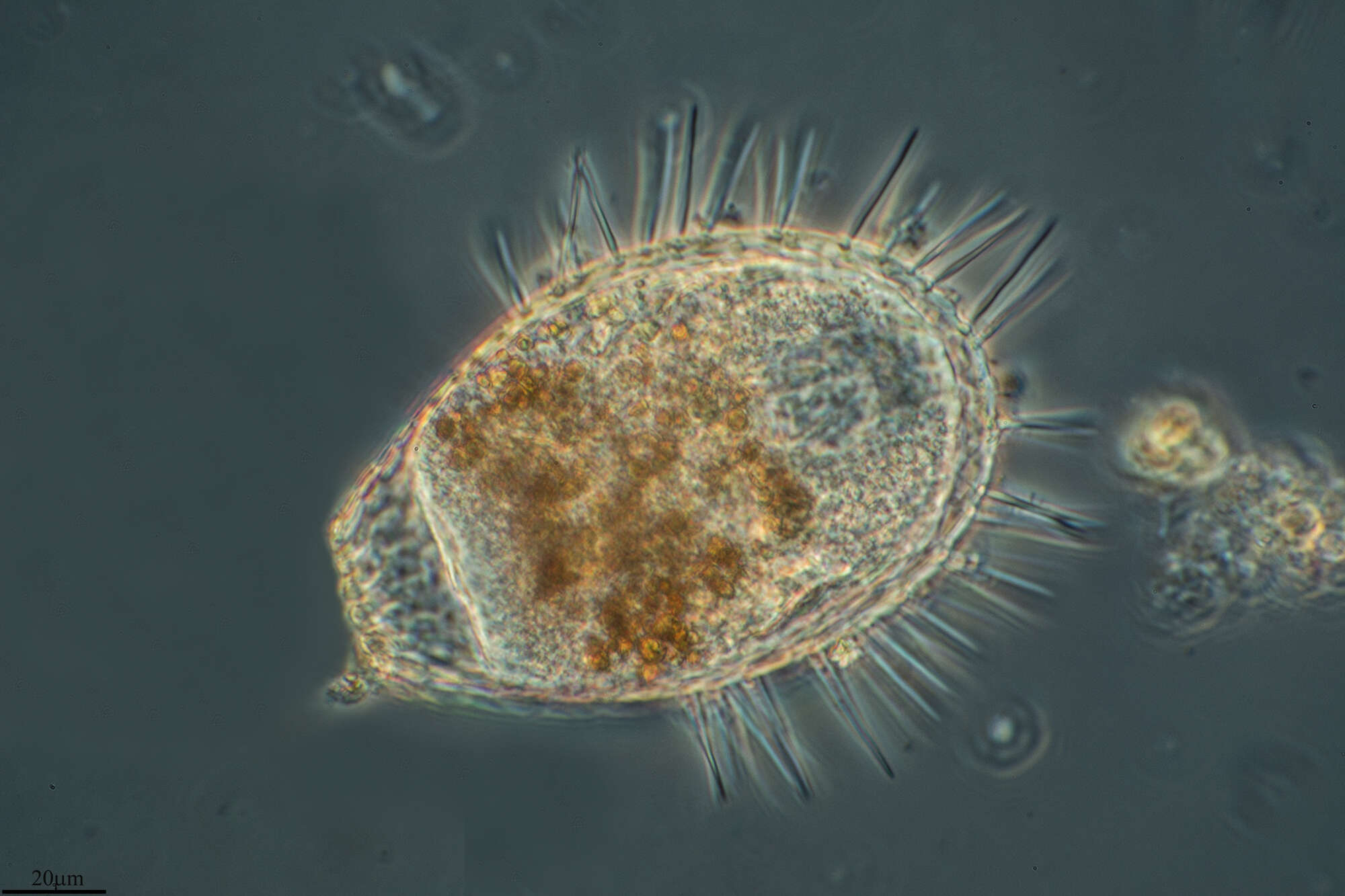
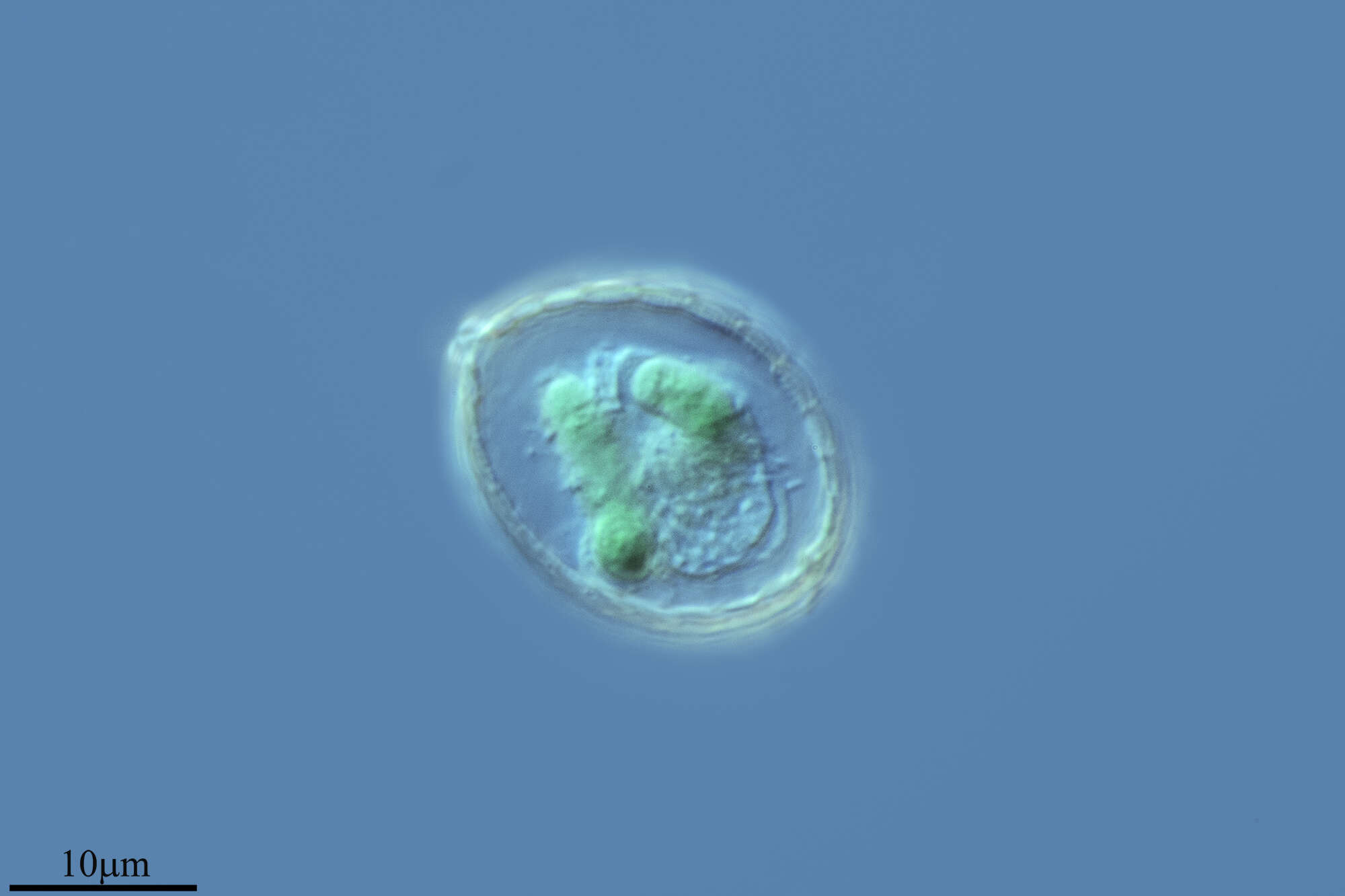
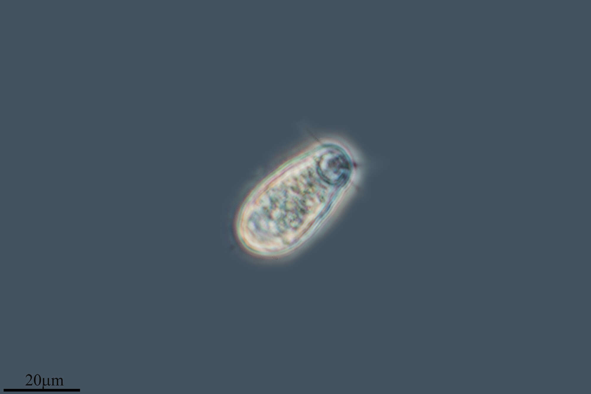
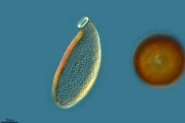
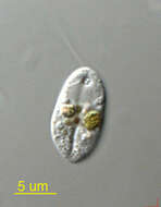
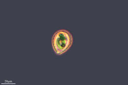
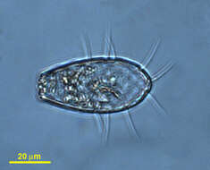
Protaspis (pro-tass-piss) obliqua Larsen and Patterson, 1990. Cells are slightly oval or roundish, 8 to 32 microns long, 10 to 27 microns wide, dorso-ventrally flattened and with thickened cortex. There is a ventral median groove, cell indented anteriorly and posteriorly where the groove meets margin. Subapically, the right margin of the groove forms a protrusion. With two flagella inserting under the protrusion, the anterior flagellum is about 0.5 times the length of the cell and the posterior flagellum is about 0.5 to 1.5 times the length of the cell. The nucleus is without nuclear caps, is located subapically in a median position, is rounded and is 5 to 13 microns in diameter. The cells may contain food particles or diatom up to 24 microns long. Commonly observed.
-
Barrio Ballinas, Castille and Leon, Spain
-
Canencia, Madrid, Spain
-
Samples from Sediment at Cedar swamps, Woods Hole, Massachusatts. Photographed by Hwan Su Yoon.