Stemonitis_lignicola_outher_net_3M
Description:
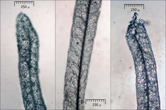
Stemonitis lignicola Nann.-Bremek, syn.: Stemonitis splendens MorenoSlo.: no name.Dat.: July 06. 2016Lat.: 46.36002 Long.: 13.70436Code: Bot_984/2016_DSC3464Habitat: grassy yard of a farmhouse, open place; almost flat terrain, calcareous, skeletal ground; partly in shade; partly protected from direct rain by the trough, average precipitation ~ 3.000 mm/year, average temperature 7-9 deg C, elevation 570 m (1.870 feet), alpine phytogeographical region. Substratum: eleven years old, handmade wooden water trough (Larix decidua wood), standing on four thin wooden legs; wood still (almost) intact, the trough still capable to hold water.Place: Lower Trenta valley, between villages Soa and Trenta, right bank of river Soa, next to Trenta 2, Skokar (abandoned) farm house, East Julian Alps, Posoje, Slovenia EC. Comment: Spore dimensions and type of surface, size and color of the sporocarps, small meshed, persistent peridial net, sparsely branched internal capillitium with extensions, wavy columella toward its end, columella reaching the apex of the sporocarps, as well as habitus and substratum fit well to the description of Stemonitis lignicola in the literature. Sporocarps 8.2-8.5 mm high, stalks 2- 2.1 mm long. Spore print abundant, dark brown. What I am curious is, did the whole development process of this myxomicete from start to the end take place on the water trough (which is most of the time dry and standing in free air), or did this thing really climb up one of the thin trough's legs in the form of plasmodium before building sporocarps?Spores evenly, densely and minutely warty (verruculose); globose to subglobose. Dimensions: 6,8 [7,2; 7,4] 7,8 x 6 [6,6 ; 6,8] 7,3 microns; Q = 1 [1,1] 1,2; N = 40; C = 95%; Me = 7,3 x 6,7 microns; Qe = 1,1. Taken from dried material by gently tapping sporocarps. Outer net (peridium) mesh dimensions: 7.5 [19.3 ; 23.2] 35 x 7 [15.3 ; 18] 26.3 microns; Q = 0.8 [1.2 ; 1.4] 1.8; N = 50; C = 95%; Me = 21.2 x 16.7 microns; Qe = 1.3. The largest meshes haven't been taken into account; assuming they are the consequence of damages during microscopy. Olympus CH20, NEA 100x/1.25, magnification 1.000 x, oil (spores), in water; NEA 40x/0.65, magnification 400x (capillitium extensions), NEA 10x/0.25, magnification 100x (stalk); Bausch & Lomb, 4x/0.10, magnification 40x (peridium, capillitium), in air without cover glass. AmScope MA500 digital camera. Herbarium: Mycotheca and lichen herbarium (LJU-Li) of Slovenian Forestry Institute, Vena pot 2, Ljubljana, Index Herbariorum LJFRef.:(1) B. Ing, The Myxomycetes of Britain and Ireland,The Richmond Publ. Co.Ltd, (1999), p 203. (2) M. Poulain, M. Meyer, J. Borronet, Les Myxomycetes, FMBDS (2011), Vol.1., p 541. (3) M. Poulain, M. Meyer, J. Borronet, Les Myxomycetes, FMBDS (2011), Vol.2., p 538. (4) Personal communication with Mr. Marko Sovre, owner of the water trough, who showed me the find.
Included On The Following Pages:
- Life (creatures)
- Cellular (cellular organisms)
- Eukaryota (eukaryotes)
- Amoebozoa (amoeboid protists)
- Evosea
- Eumycetozoa (slime molds)
- Myxogastria (plasmodial slime molds)
- Columellidia
- Stemonitida
- Stemonitidae
- Stemonitis
- Stemonitis lignicola
This image is not featured in any collections.
Source Information
- license
- cc-by-nc-sa
- copyright
- Amadej Trnkoczy
- photographer
- Amadej Trnkoczy
- original
- original media file
- visit source
- partner site
- Flickr Group
- ID