Trametes-pubescens_spore_1M

Description:
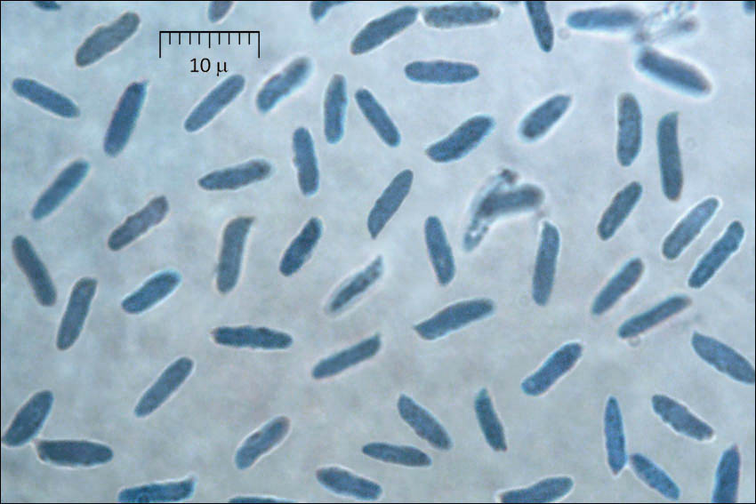
Trametes pubescens (Schumach.: Fr.) Pilt, syn.: Coriolus pubescens (Schumach.: Fr.) Murr.DE: Samtige TrameteSlo.: puhasta ploskocevkaDat.: Nov. 17. 2015Lat.: 46.40365 Long.: 13.74211Code: Bot_927/2015_DSC9883Habitat: mixed wood, Fagus sylvatica, Picea abies, dominant trees with some Larix decidua and Fraxinus excelsior; steep mountain slope, west aspect, however in shade of mountains during winter months; rather cool and humid place; calcareous ground; partly protected from direct rain by tree canopies; average precipitations ~ 3.000 mm/year, average temperature 5 - 6 deg C, elevation 790 (2.600 feet), alpine phytogeographical region.Substratum: large, very old Fagus sylvatica, the tree is still alive, but fungus is growing on partly dead part of it, still in bark.Place: Zadnja Trenta valley; at the border of Forest reserve Kukla; about 100 m southwest of the memorial of Dr. Julius Kugy, poet and mountaineer of Julian Alps; near switchback no.48 of Vri alpine road, East Julian Alps, Posoje, Slovenia EC.Comments: This find has posed very interesting challenges for determination. Several options have been considered but apparently none fit to the find. Experts have been consulted but no definite solution found. Finally a sample has been sent to Dr. Leif Ryvarden, University of Oslo who determined it as Trametes pubescens. Many thanks to all involved in the problem solving - Dr. Nikica Ogris and Andrej Piltaver, Slovenian Forestry Institute, Bojan Rot, Bovec, Branko Vrhovnik, Horjul and specially to Prof. Ryvarden for his final determination.The main source of identification problems is probably very untypical shape of the pilei. Trametes pubescens has usually relatively thin pilei. According to the key of genus Trametes given in Krieglsteiner (2000), p585 the pilei are about 0.5 (1) cm thick, Ryvarden (2014), p417 states: "... Basidiocarps thin ... context up to 5 mm ... pore layer up to 4 mm thick..." and Bernicchia (2005), p535 gives " ... context 3-5 mm and pore layer 1-5 mm thick... ". The pilei found were up to 5 cm thick and in most cases triquetrous in cross-section. Also spores are significantly longer than normally expected.Growing in a few groups on the same part of a large tree; altogether more than 200 pilei present; majority of them laterally confluent, imbricate, some single; some effuse-reflexed, most of them triquetrous in cross-section; pilei dimensions: 8-10(16) x 4-5(6.5) cm and 2.5-5 cm thick; pore layer up to 8 mm thick; context of very low specific weight, corky, similar to dry Piptoporus betulinus; when dry quite firm, brittle, brakes to pieces; smell (of almost dry pilei) very mild but distinctive on what? ; taste indistinctive at the beginning, after a while mild and interesting, again on what?; 5% KOH reaction on context and pileus surface yellow-ocher with orange tint, on pores the same color but less distinctive; SP scarce, but distinctive (after making pilei moist and at 18-20 deg C), whitish-beige, oac851; fungi causing white root according to analysis of the wood made at the Forestry Institute of Slovenia.Spore dimensions determined twice from SP of different pilei. First measurement (pilei taken on Nov. 18. 2015): 7 [7.8; 8] 8.8 x 2 [2.4; 2.5] 2.9 microns; Q = 2.6 [3.2; 3.4] 3.9; N = 49; C = 95%; Me = 7.9 x 2.4 microns; Qe = 3.3. Second measurement (pilei taken on Nov. 23. 2015): 6.1 [7.3; 7.6] 8.9 x 2 [2.3; 2.4] 2.8 microns; Q = 2.5 [3.1; 3.2] 3.9 ; N = 62 ; C = 95%; Me = 7.5 x 2.4 microns; Qe = 3.2. Basidia clavate, dimensions: 14.2 [15.8; 17.7] 19.4 x 4.1 [5; 6.1] 7 microns; Q = 2.3 [2.8; 3.3] 3.8; N = 8; C = 95%; Me = 16.8 x 5.5 microns; Qe = 3.1. Hyphal system trimitic. Olympus CH20, NEA 100x/1.25, magnification 1.000 x, oil, in water, aniline blue; in vivo. AmScope MA500 digital camera.Herbarium: Mycotheca and lichen herbarium (LJU-Li) of Slovenian Forestry Institute, Vena pot 2, Ljubljana, Index Herbariorum LJFRef.:(1) Id'ed by Dr. Leif Ryvarden, University of Oslo.(2) G.J. Krieglsteiner (Hrsg.), Die Grosspilze Baden-Wrttembergs, Band 1., Ulmer (2000), p 589.(3) L. Ryvarden, I. Melo, Poroid fungi of Europe, Synopsis Fungorum 31., Fungiflora (2014), p 417. (4) A. Bernicchia, Polyporaceae s.l., Fungi Europaei, Vol. 10., Edizioni Candusso (2005), p 535. (5) S. Buczacki, Collins Fungi Guide, Collins (2012), p 509.
Included On The Following Pages:
- Life (creatures)
- Cellular (cellular organisms)
- Eukaryota (eukaryotes)
- Opisthokonta (opisthokonts)
- Nucletmycea
- Fungi (mushrooms, lichens, molds, yeasts and relatives)
- Dikarya
- Basidiomycota (basidiomycete fungi)
- Agaricomycetes (Mushroom-Forming Fungi)
- Polyporales
- Polyporaceae (bracket fungi)
- Trametes
- Trametes pubescens
This image is not featured in any collections.
Source Information
- license
- cc-by-nc-sa
- copyright
- Amadej Trnkoczy
- photographer
- Amadej Trnkoczy
- original
- original media file
- visit source
- partner site
- Flickr Group
- ID