-
Jaime Gonzalez-Cueto, Sigmer Quiroga, Jon Norenburg
Zookeys
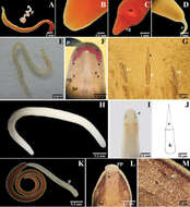
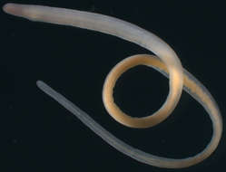
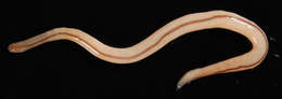
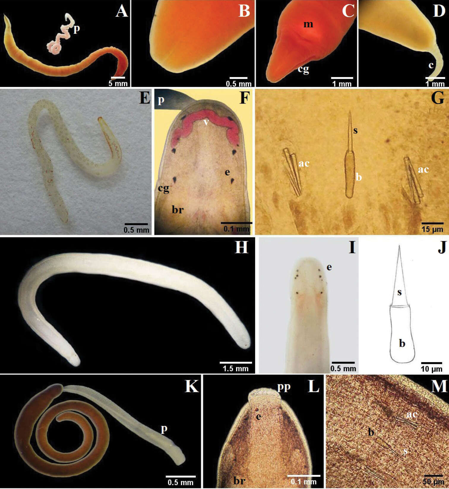
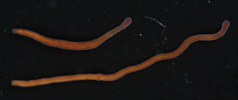
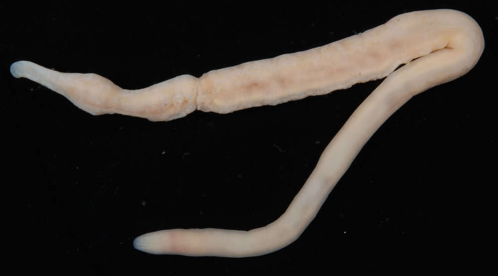
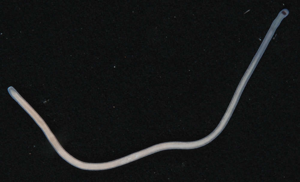
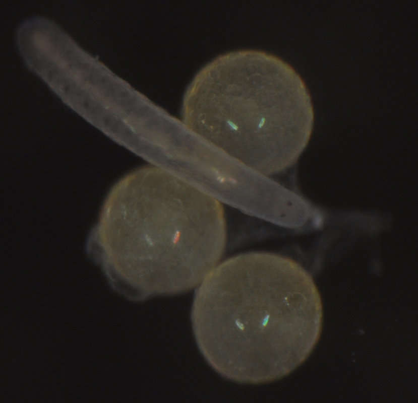
Figure 3.A–D Micrura ignea: A entire specimen, the worm has expulsed the proboscis B dorsal detail of the head C ventral detail of the head D detail of the tail E–G Amphiporus cruentatus: F dorsal detail of the head G detail of the stylets H–J Amphiporus cf. ochraceus: I dorsal detail of the head J drawing of the stylet K–M Amphiporus texanus: K entire worm L dorsal detail of the head M detail of the stylets. ac accessory stylet, b base of the stylet, br brain, c cirrus, cg cephalic grooves, e eyes, m mouth, p proboscis, pp proboscis pore, s sylet, v blood vessel.
-
Jaime Gonzalez-Cueto, Sigmer Quiroga, Jon Norenburg
Zookeys
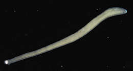
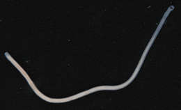
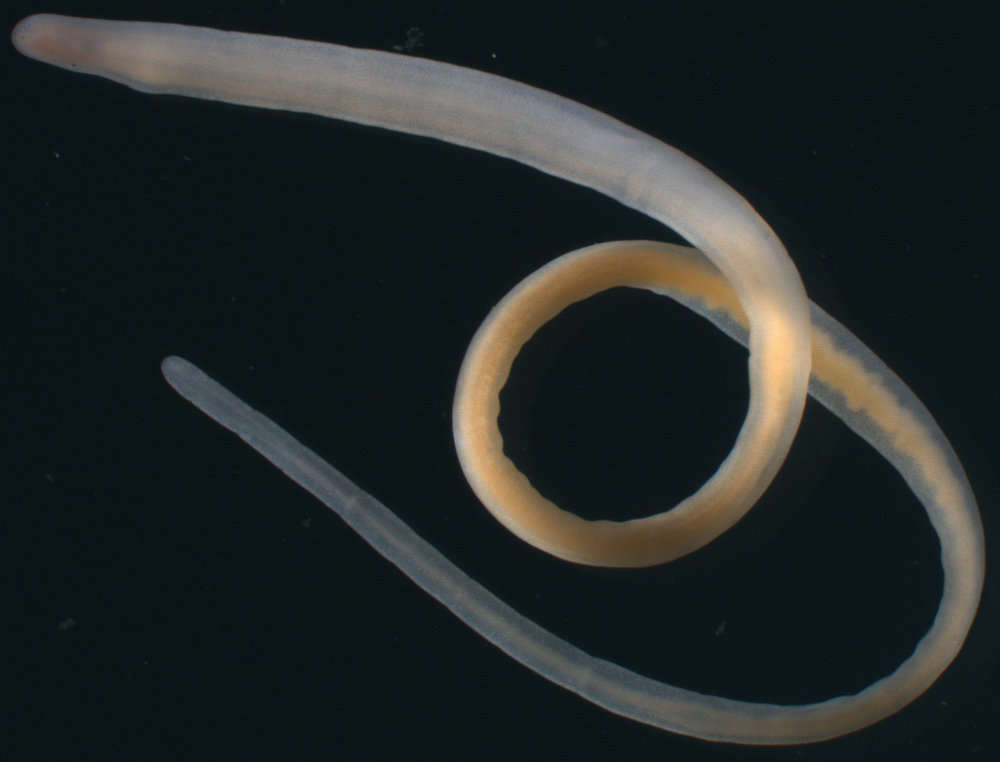
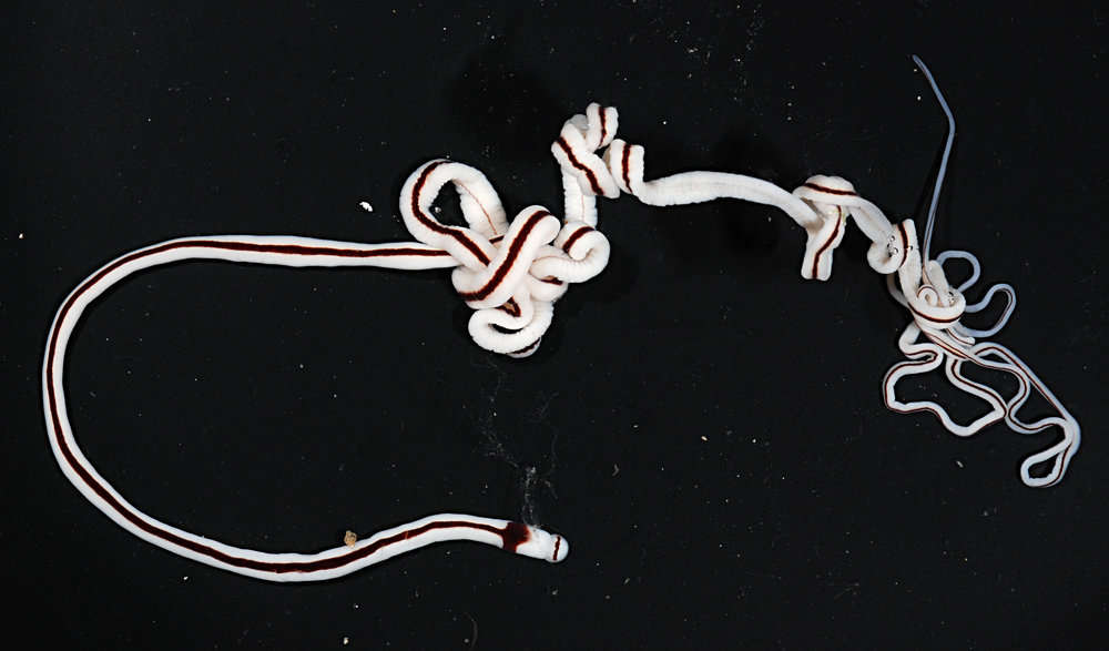
Figure 4.A–C Nemertopsis bivittata: B dorsal detail of the head and proboscis, C detail of the stylets D Ototyphlonemertes erneba (modified from Corrêa 1950 and Kirsteuer 1977) E–G Ototyphlonemertes lactea: E entire worm F detail of the statocysts G detail of the stylets H–J Zygonemertes fragariae: H entire worm I detail of the head J detail of the stylets K–M Zygonemertes virescens: K entire worm L detail of the head M detail of the stylets. ac accessory stylet, b base of the stylet, br brain, c sensorial cirrus, e eyes, p proboscis, pa proboscis papilla, s sylet, st statocysts.
-
All Biocode files are based on field identifications to the best of the researcher’s ability at the time.
-
All Biocode files are based on field identifications to the best of the researcher’s ability at the time.
-
All Biocode files are based on field identifications to the best of the researcher’s ability at the time.
-
All Biocode files are based on field identifications to the best of the researcher’s ability at the time.
-
All Biocode files are based on field identifications to the best of the researcher’s ability at the time.
-
All Biocode files are based on field identifications to the best of the researcher’s ability at the time.
-
All Biocode files are based on field identifications to the best of the researcher’s ability at the time.
-
All Biocode files are based on field identifications to the best of the researcher’s ability at the time.
-
All Biocode files are based on field identifications to the best of the researcher’s ability at the time.
-
All Biocode files are based on field identifications to the best of the researcher’s ability at the time.
-
All Biocode files are based on field identifications to the best of the researcher’s ability at the time.
-
All Biocode files are based on field identifications to the best of the researcher’s ability at the time.
-
All Biocode files are based on field identifications to the best of the researcher’s ability at the time.
-
All Biocode files are based on field identifications to the best of the researcher’s ability at the time.
-
All Biocode files are based on field identifications to the best of the researcher’s ability at the time.
-
All Biocode files are based on field identifications to the best of the researcher’s ability at the time.
-
All Biocode files are based on field identifications to the best of the researcher’s ability at the time.
-
2011 California Academy of Sciences
CalPhotos
-
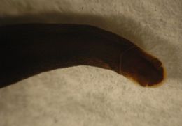
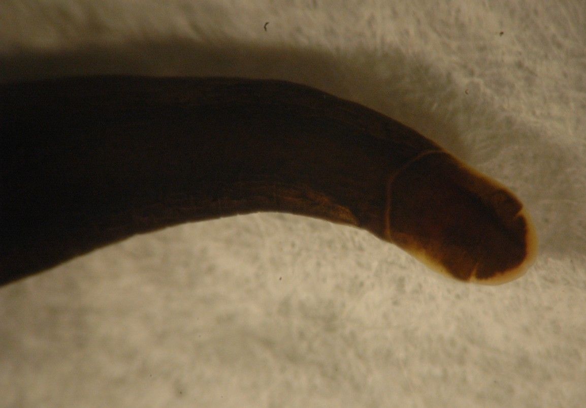
This view of the head shows the lighter colored band along the edge of the head and across the back of the head. The most posterior clusters of ocelli are near the posterior end of the band along the border.
-
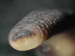
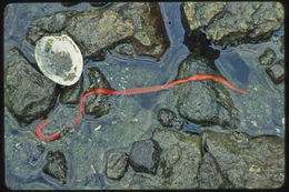
This view of the front of the head (contracted, alcohol-preserved specimen) shows the combined mouth and proboscis at the front of the head. Photo by Dave Cowles, March 2010