Portrait
Description :
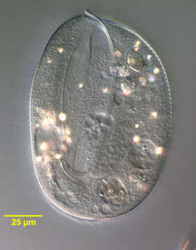
Portrait (optical section) of the oligohymenophorean ciliate, Lembadion bullinum (Müller,1786;Perty,1849). Cell outline is oval. The ventral surface is concave and the dorsum convex. The very large scoop-like peristome occupies most of the ventral surface. The cytostome is at the posterior end of the peristome. There is a small undulating membrane on the right margin of the peristome. A large sheet-like adoral membranelle arises from the left margin of the peristome. There are 40-60 evenly spaced longitudinal somatic kineties. The pellicle of L. bullinum, divided into small roughly rectangular depressions, has a distinct cribriform pattern. This featurte distinguishes L. bullinum from L. magnum which has a striate pellicular pattern. There is usually a tuft of longer caudal cilia. The contractile vacuole (seen just anterior to the macronucleus here) connects with its excretory pore by a long curved canal (not seen here). The single ovoid macronucleus (seen here) and micronucleus (not seen in this image) are posterior. Collected from a freshwater pond near Boise, Idaho May 2004. DIC optics.
Inclus dans les pages suivantes :
- Life
- Cellular (Organismes cellulaires)
- Eukaryota (eucaryotes)
- SAR (Stramenopiles, Alveolates, Rhizaria)
- Alveolata
- Ciliophora
- Intramacronucleata
- Oligohymenophorea
- Peniculida
- Lembadionidae
- Lembadion
- Lembadion bullinum
Cette image ne figure dans aucune collection.
Informations sur la provenance
- licence
- cc-by-nc
- auteur
- William Bourland
- fournisseur
- micro*scope
- original
- fichier de média d’origine
- visiter la source
- site partenaire
- micro*scope
- ID